Dortmund, 9th June 2026
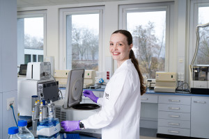
Inside the laboratory, the atmosphere is one of intense concentration: intern Leonie Menzel and her academic supervisor, Dr Malte Roeßing, are calibrating the light sheet fluorescence microscope. Using high-resolution 3D imaging, they aim to examine a small section of a human coronary artery. A potentially dangerous atherosclerotic plaque has formed in this blood vessel. To find out why, the two researchers want to look at cellular changes.
The light sheet fluorescence microscope spreads the laser beam out like a sheet of paper. The resulting thin sheet of light illuminates every single layer of the sample – and captures an image of each layer. The researchers will later assemble the individual images on a computer to create a three-dimensional model. The images taken today produce a 3D image of the coronary artery, including plaque, in which the two biologists can now analyse the spatial distribution of individual fluorescently labelled cells throughout the tissue.
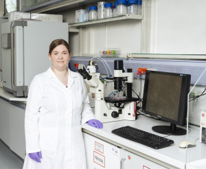
Leonie Menzel at the light sheet fluorescence microscope. During her internship, she spends a lot of time in the laboratory, steadily expanding her methodological knowledge and refining the research question for her Master’s thesis.
© ISAS
An unusual view into the coronary artery
For Menzel, it is the first time she is seeing a human coronary artery under a light sheet fluorescence microscope. The 26-year-old is studying medical biology at the University of Duisburg-Essen. At ISAS, she is completing a six-week internship as part of her master’s degree. On the screen in front of her, the coronary artery appears as a 3D reconstruction. By rotating the sample, Menzel can examine the tissue, including the plaque, from different perspectives with high precision.
“Typically, plaques form on curved or branching sections of blood vessels. A well-known cause of plaques is the mechanical effect of blood flow on the vessel wall,” explains Roeßing, a postdoctoral researcher in the Bioimaging research group. “However, plaques can also occur on straight, unbranched sections. The underlying mechanisms here are still largely unclear.” Plaques develop as a result of cellular remodelling processes in the arterial wall. The accumulation of fats there triggers an immune response, causing immune cells to migrate into the vessel wall. Over time, these can narrow the vessels – with potentially serious consequences such as heart attacks or strokes.
Internship: sample preparation for imaging
“I find it fascinating to investigate the causes of diseases at a molecular level,” says Menzel. Specifically, she and Roeßing are examining changes in the cellular composition of the various layers of the coronary artery. To make this possible, Menzel prepared the sample beforehand with the assistance of postdoc Roeßing. To do this, she treated the section of vessel with, among other things, cinnamic acid ethyl ester – a process known as ‘clearing’, which makes the tissue sample optically transparent. The cell structures scatter and absorb very little light, allowing the laser beam to pass through the sample.
In addition, the early-career researcher labelled the tissue with fluorophore-conjugated antibodies. The antibodies recognise structures on or within cells. When the laser excites the fluorophores at the appropriate wavelength, they emit light at a characteristic wavelength (see info box). In this way, Menzel is able to localise individual cells, such as endothelial cells lining blood vessels or macrophages (the immune system’s scavenger cells), within the vessel wall. This makes it possible to analyse disease-related changes in cell distribution. In this case, the aim is to gain a better understanding of the cellular mechanisms underlying plaque formation. Menzel made a significant contribution to this work during her time at ISAS.
Fluorescent markers
Fluorophores are fluorescent molecules, such as dyes or fluorescent proteins, which bind to molecular targets on the surface or inside cells (for example, proteins, DNA, enzymes or viruses). When excited by specific wavelengths of light, the fluorophores on the labelled cellular components emit light in different colours.
Research experience for the master’s thesis
“It’s really exciting to be working in the lab as an intern, not only to learn about new analytical methods, but also to be able to put them into practice,” says Menzel of her time at ISAS. She will return to the institute to work on her master’s thesis. During her stay in Dortmund, she aims to build on her previous experimental experience, further develop her methodological skills, and gradually refine her scientific research question.
(Saskia Schlesinger)