The research group aims to detect the molecular and cellular processes that underlie so-called immuno-vascular interactions under inflammatory conditions. In order to understand, for example, an infection and its corresponding immune response, the scientists look at the process from different angles.
One of the imaging techniques that the researchers use is fluorescence microscopy. This form of light microscopy includes, among others, light sheet fluorescence microscopy (LSFM). LSFM enables the scientists to determine information on different classes of molecules and their spatial distribution patterns at the same time. In addition to imaging techniques like LSFM, the researchers also work with confocal laser scanning microscopy (CLSM) and two-photon laser scanning microscopy (TPLSM). They enable a three-dimensional analysis of biological samples from a cellular to sub-cellular level.
The researchers conduct studies on animal and human samples, take measurements of intact organs and integrate artificial intelligence into their image analyses.
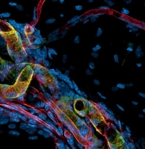
To ensure bone homeostasis, the bone surface – the periosteum – and the cortical bone are supplied by different types of blood vessels, such as fine capillaries (red) and large arteries (green and red).
© ISAS / Anika Grüneboom
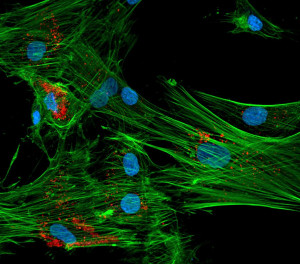
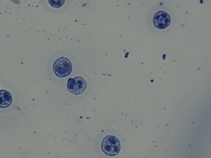
Distribution of lysosomes (red) in human mesenchymal stem cells. The cytoskeleton is shown in green and the nuclei are blue.
© ISAS / Anika Grüneboom
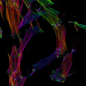
Colour coding of the spatial orientation of actin fibers in human fibroblasts.
© ISAS / Anika Grüneboom
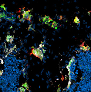
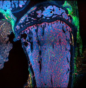
Identification of resorptively active (red, green, white) and inactive osteoclasts (green, white) at the growth plate and trabeculae.
© ISAS / Anika Grüneboom
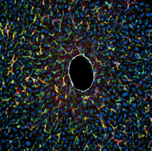
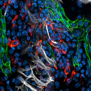
Hepatic macrophages (red) primarily interact with sinusoidal endothelial cells (green), while hardly any immune cell interactions are observed at the central vein (white).
© ISAS / Anika Grüneboom
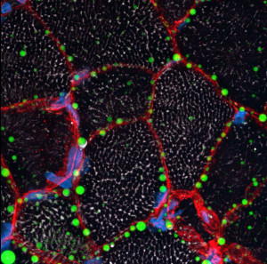
The staining of neutral lipids (green) and basement membrane (red) allows the detection of lipid droplets lined up along the endomysium of a patient with congenital myasthenic syndrome. The mitochondria (white) have lower amounts of neutral lipids than the endomysium.
© ISAS / Anika Grüneboom
Localisation of osteoclasts (green) along osseous blood vessels (red) in the murine mandible.
© ISAS / Anika Grüneboom
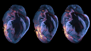
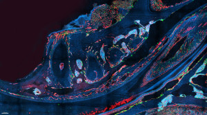
Light sheet fluorescence microscopy of immune cell infiltration (red-orange) in a murine heart (blue) after myocardial infarction.
© ISAS / Anika Grüneboom
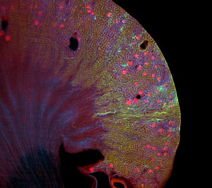
Light sheet fluorescence microscopy of the spatial distribution of different macrophage populations (blue and green) along blood vessels and glomeruli (red) in a murine kidney.
© ISAS / Anika Grüneboom
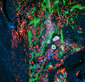
Infiltration of immune cells (red) and inflammation-driven angiogenesis of blood vessels (green and white) in a murine knee joint during rheumatoid arthritis.
© ISAS / Anika Grüneboom
Infiltration of immune cells (red) and inflammation-driven angiogenesis of blood vessels (green and white) in a murine knee joint during rheumatoid arthritis.
© ISAS / Anika Grüneboom
The blood vessel architecture of the murine tibia includes both venous sinusoids (red) and arteries (yellow) in the bone marrow, as well as arterial (yellow) and venous (red) transcortical vessels connecting the bone marrow through the cortical bone to the peripheral vasculature.
© ISAS / Anika Grüneboom