Dortmund, 12th August 2025
When researchers examine tissues in order to get to the bottom of disease mechanisms, they often need to choose between two fundamental approaches. On the one hand, they can mark substances specifically and, for example, use microscopic techniques to investigate how many of them are present in the tissue. For this quantitative analysis, however, the researchers must already know what they’re looking for. On the other hand, they can use methods such as mass spectrometry imaging to see what metabolic products, for example, are present in the tissue in the first place. Until now, however, it has been difficult to read off the quantity of the substances from this qualitative data. Now, with the participation of ISAS, a group of researchers has refined a method that allows not only qualitative but also quantitative analysis – and, for the first time, for a whole class of substances.
To this end, the team led by Prof. Dr Bernhard Spengler from Justus Liebig University Giessen and Prof. Dr Sven Heiles, leader of the Lipidomics research group at ISAS, have combined two analytical techniques known as “atmospheric-pressure matrix-assisted laser desorption/ionization mass spectrometry imaging” (AP-SMALDI MSI) and “nanoflow hydrophilic-interaction liquid chromatography tandem mass spectrometry” (nano-HILIC MS/MS). Using this combined method, they investigated the molecular processes taking place in schistosomiasis, a neglected tropical disease with over 200 million sufferers worldwide. The researchers published their results in the journal Analytical Chemistry.
Molecular insights into schistosomiasis
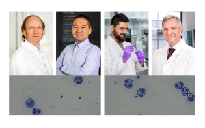
Schistosomiasis, also known as bilharzia, is triggered when people come into contact with water contaminated with the larvae of Schistosoma worms. These tiny parasites enter the body by penetrating the skin and lay eggs that are often deposited in the organs – especially the liver. The human immune system responds by forming granulomas (spherical tissue structures) around the eggs. These structures are actually intended to encapsulate the parasites to prevent their spread. However, this defensive response often leads to inflammation, which can cause conditions including fibrosis – that is, chronic scarring – of the liver. Until now, it has been difficult to determine what this process entails on the molecular level.
In their study, the researchers focused on a specific group of fats known as glycosphingolipids (GSLs). These glycolipids, which are a key constituent of the cell membrane, are structured like a lollipop: with a fat-soluble “backbone”, known as a ceramide, which is anchored in the membrane, and a water-soluble sugar headgroup that projects outwards. GSLs play a key role in the communication between cells and in immune responses, acting as “immunomodulators”: antibodies, endogenous cells and immune cells detect GSLs and produce an immune response accordingly. So far, however, there has been no way of determining how many of these molecules are present in a specific tissue using imaging.
Article Recommendation
Luh, D., Heiles, S., Roderfeld, M., Grevelding, C.G., Roeb, E., Spengler, B.
(2024) Hepatic Topology of Glycosphingolipids in Schistosoma mansoni-Infected Hamster. Analytical Chemistry, 96(16):6311-6320.
Finally, chemical imaging with relative quantities
In their method, the team led by Spengler and Heiles combined the chemical analysis of nano-HILIC MS/MS with the high-resolution imaging of AP-SMALDI MSI in order to examine the liver tissue of healthy hamsters and hamsters infected with Schistosoma. In AP-SMALDI MSI, a fine laser beam scans the tissue point by point and breaks off molecules, which are identified by the mass spectrometer. This produces a chemical image of the tissue, resembling the image produced by a thermal imaging camera but showing the spatial distribution of certain molecules rather than temperatures. By optimising the pixel resolution of the AP-SMALDI MSI method to as little as three micrometres – about a 20th of the thickness of a human hair – the scientists were able to recognise even the finest structures within the granulomas and therefore to distinguish between antigens and endogenous GSLs, for example, in the case of the parasitic infection.
The key advance is that, with their method, the researchers were able not only to see where GSLs were located in the animals’ tissue but also to compare the relative amounts in different tissue regions – an important step that goes beyond just imaging. Specifically, the team identified 60 different GSL species and established that 50 of them occurred in greater quantities in infected tissue. Of these molecules, 44 were directly connected with schistosomiasis-related granuloma formation.
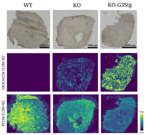
Lipid deposits in the heart in a mouse model for Fabry disease: The image shows heart tissue from mice in three groups: on the left, the tissue is from healthy wild type (WT) animals. In the middle, it is from knock-out (KO) mice in which the gene for the enzyme α-galactosidase A has been specifically switched off. This enzyme normally breaks down glycosphingolipids (GSLs) but is diminished in people with Fabry disease. In the right column are the samples from knock-out mice that also produce greater quantities of GSLs (K0-G3Stg), exacerbating the disease characteristics. The top row shows the tissue under an optical microscope. The middle and bottom rows each show the same sections using mass spectrometry imaging: in the middle row, the disease-relevant GSL Gb3Cer 34:1 is highlighted in colour, while the bottom row shows the cell membrane lipid PC 34:1, which occurs in the normal state, for comparison. The colour scale of the measurement results from minimum to maximum intensity clearly shows that the stronger the typical Fabry-disease conditions, the greater the quantity of harmful GSLs deposited in the heart tissue.
© Sven Heiles
More than just an image – a paradigm shift
This work represents a paradigm shift, moving away from purely qualitative imaging (“what is where?”) to quantitative analysis (“how much is where?”). The developed method not only opens up new insights into the pathology of schistosomiasis but also has potential applications in various areas of biomedicine where the spatial distribution of lipids and other biomolecules plays a significant role. At ISAS, for example, Heiles and his team are working on analytical techniques for use in a genetic lipid storage disorder known as Fabry disease. In patients, this disorder leads to an accumulation of GSLs that is harmful in the long term. The ability to better understand and analyse this process could potentially pave the way for new therapies.
(Ute Eberle)