Skip to content

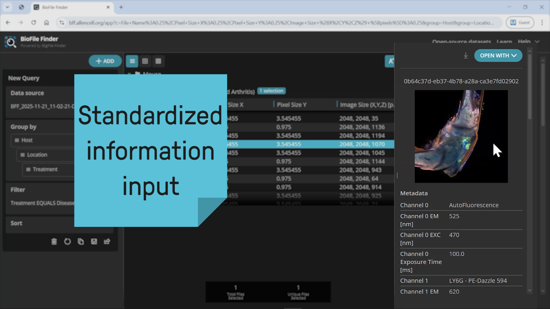
The aim of the junior research group AMBIOM – Analysis of Microscopic BIOMedical Images, which is funded by the Federal Ministry of Research, Technology and Space (Bundesministerium für Forschung, Technologie und Raumfahrt, BMFTR), is to enable a high analytical throughput of microscopic images. The research group plans to develop algorithms and methods (open source) by 2026 that will allow countless image data worldwide to be analysed automatically, quickly and economically.

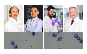
© ISAS / Hannes Woidich

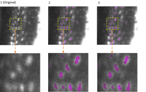
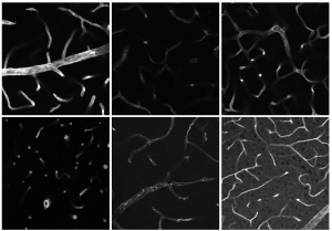
Focus: large 3D microscopy imaging data

The research group concentrates on the development of scalable AI-based biomedical image analysis algorithms, particularly for large 3D microscopy image data. AMBIOM’s work aims to allow broad-based new studies on the development of diseases and their consequences at the level of entire organs and organ systems. Furthermore, the AI analysis methods developed at ISAS are intended to help doctors in making diagnostic and therapeutic decisions.

The MSCoreSys associated junior research group AMBIOM – Analysis of Microscopic BiOMedical Images is funded by the Federal Ministry of Research, Technology and Space (Bundesministerium für Forschung, Technologie und Raumfahrt, BMFTR) under the funding reference 161L0272.

Highlights

PhD Candidate (m/f/d)

24th January 2024

EfficientBioAI: New Open-Source Software Makes AI Models Lighter and Greener

Artificial intelligence has become an integral part of research. However, the better and more complex the models become, the higher their energy consumption. Researchers at ISAS and Peking University have therefore developed ready-to-use and open software that compresses existing bioimaging AI models. With the help of the new toolbox, scientists can now run their models faster and with significantly lower energy consumption.

Die Abbildung zeigt die 3D-Semantiksegmentierung von Osteozyten in Mäuseknochen vor und nach der Kompression des Modells mir EfficientBioAI Die Bilder wurden mittels Lichtblatt-Fluoreszenzmikroskopie aufgenommen.
2nd July 2026

Biomedical Image Analysis with AI

Dr Jianxu Chen is taking up a professorship in AI for Biomedical Analysis at the University of Duisburg-Essen. With his research group at ISAS, he is developing AI methods that can be used to comprehensively analyse enormous quantities of complex microscopy images.

Portrait von Dr. Jianxu Chen.
29th June 2026

It's What's Inside That Counts: Neutrophils Form Distinct Subtypes

Neutrophils can be divided into two distinct subgroups with completely different functions. The surface protein CD177 plays a decisive role in this. For the first time, researchers from the Faculty of Medicine at the University of Duisburg-Essen and ISAS have been able to demonstrate, using a new integrated proteomics and lipidomics analysis, that the molecular signatures in CD177-defined neutrophils differ significantly from one another. The presence of this surface protein has implications for disease progression, for example in head and neck tumours.

CD177 negative Neutrophile.
2nd July 2026

Four perspectives on a shared discovery

Although CD177⁺ and CD177⁻ neutrophils look identical under the microscope, they are in fact not the same. Four of the authors of “CD177 Deficiency Defines a Stable Subtype of Human Neutrophil Granulocytes with Tumour-Promoting Activity” (Advanced Science) explain how they identified the functional differences between these immune cells and which combined analytical methods made their discovery possible in the first place. This insight into their research demonstrates how interdisciplinary collaboration can reveal previously undiscovered information.

Das Foto zeigt eine Collage mit Porträts von vier Personen und Mikroskopaufnahmen der Immunzellen Neutrophilen Granulozyten; Von oben links nach rechts: Prof. Dr. Matthias Gunzer, Prof. Dr. Jianxu Chen, Dr. Belal Alshaar und Prof. Dr. Albert Sickmann.
28th May 2026

3 Questions for Lukas Fu

How can AI support the analysis of biomedical images? This was the question Lukas Fu explored during his school internship at ISAS. The 15-year-old also successfully completed his first programming project.

Lukas Fu, a 15-year-old with short black hair. He is wearing glasses and is sitting on a low wall.
13th May 2026

Separating Fluorescence Signals Faster and More Precisely with AI

When different fluorescence signals overlap during microscopy, their clear assignment to specific biological structures becomes difficult. To address this common problem, Dr Davide Panzeri is developing AI-based signal separation methods. The biophysicist has been awarded a Marie Skłodowska-Curie Fellowship by the European Union for his promising research project.

Porträt Dr. Davide Panzeri.
17th April 2026

ISAS BFF Uploader

13th October 2025

New Framework for Efficient Image Data Analysis in Biomedicine

How can AI be used optimally in bioimaging research? A team of international researchers and scientists led by Dr Jianxu Chen from ISAS has developed a framework designed to make it easier for biomedical scientists to work with data-centric AI. Using the example of vascular segmentation, the authors of the publication in the journal npj imaging now demonstrate the advantages of the new framework.

30th April 2025

Humboldt Fellow Prof Dr Xiaowei Xu started at ISAS in March

Prof Dr Xiaowei Xu from the Chinese Guangdong Cardiovascular Institute is researching the clinical application of artificial intelligence (AI) in the context of cardiovascular diseases. For 18 months in total, he will be researching various AI methods for analysing cell images as a Humboldt Fellow at ISAS.

Prof Dr Xiaowei Xu in front of the ISAS city building.
27th March 2025

Valuable Connections: Dr Jianxu Chen

Dr Jianxu Chen leads the junior research group AMBIOM - Analysis of Microscopic BIOMedical Images at ISAS since 2021. He previously worked at the Allen Institute for Cell Science in Seattle, USA. In the ISAS -Kompakt series ‘Valuable Connections’, the computer scientist reports on his move to health research and to Germany.

Portrait von Dr. Jianxu Chen.
26th February 2025

What are you doing at ISAS, Leon?

What do marshmallows and chocolate have to do with cell analysis? Leon is finding out the answer during his school internship at ISAS. For ISAS Kompakt, the 15-year-old talks about what else he is learning during his time at the institute.

Leon hält Marshmallows, Schokolade und die Hardware für sein Projekt zur Bilderkennung.
4th February 2025

Valuable Connections: Adrian Sebuliba

Adrian Sebuliba joined the ISAS junior research group AMBIOM in 2023 as a software engineer. Previously, he worked for a digital commerce platform for the chemical industry in Uganda. In the ISAS Kompakt series ‘Valuable Connections’, he reports on his move into health research, among other things.

Portrait of Adrian Sebuliba.

Projects

AI Assisted Imaging of Large Tissues

Researchers on the »Imaging of Large Tissues« project are developing a workflow to combine the various microscopy imaging methods and analytical, mass spectroscopy methods.

Biochemical Annotations of Mass Spectrometry Imaging Data for Worldwide Exchange

The research groups AMBIOM and Spatial Metabolomics are working together to develop a plug-in for the multi-dimensional imaging software napari that makes it possible to visualise and biochemically annotate MSI data.

Creating 'Leibniz Mass Spectral Imaging Library' for Identification of Primary & Secondary Metabolites

The project aims at creating the first-ever open-access MSI library of over 1000 bioactive compound standards on different matrix-assisted laser desorption/ionization (MALDI) MSI platforms.

Smart Human-in-the-loop Segmentation

Scientist working on the project »Smart Human-in-the-loop Segmentation« aim to develop a powerful deep learning model that is trained with a minimum amount of human effort.

TRR 332 – Neutrophils: Origin, Fate & Function

The DFG Collaborative Research Centre Transregio 332 is investigating the role of neutrophils, a type of immune cell, in various diseases. ISAS is involved in this project through two sub-projects, with the long-term aim of enabling new therapeutic approaches, for example for rheumatoid arthritis.

Cell Tracking in Microscopy Images

The research project »Cell Tracking in Microscopy Images« aims to develop new sophisticated cell detection and tracking algorithms to tackle some of the most challenging tracking problems.

Team

Dr. Jianxu Chen

Research Group Leader

Department: Biospectroscopy

Research group: AMBIOM

Portrait von Dr. Jianxu Chen.

Yin Hang

Research Associate

Department: Biospectroscopy

Research group: AMBIOM

Lukas Johanns

Research Associate

Department: Biospectroscopy

Research group: AMBIOM

Portrait von  Lukas Johanns.

Lennart Kowitz

IT system administrator

Department: Biospectroscopy

Research group: AMBIOM

Portrait von  Lennart Kowitz.

Dr. Davide Panzeri

Research Associate

Department: Biospectroscopy

Research group: AMBIOM

Portrait von  Davide Panzeri.

Simon Püttmann

Research Associate

Department: Biospectroscopy

Research group: AMBIOM

Portrait von  Simon Püttmann.

Justin Sonneck

Research Associate

Department: Biospectroscopy

Research group: AMBIOM

Portrait von  Justin  Sonneck.

Jonathan Jair Sánchez

Research Associate

Department: Biospectroscopy

Research group: AMBIOM

Portrait von  Jonathan Jair Sánchez.

Yu Zhou

Research Associate

Department: Biospectroscopy

Research group: AMBIOM

Portrait von  Yu Zhou.