Most illnesses or medical conditions, including cardiovascular diseases or tumours, are associated with localised and heterogeneous changes of biochemical cascades within some cells. The reason is that cells often respond differently when triggered with external stimuli such as low oxygen levels, viruses, bacteria, or when genetic alteration occur. This leads to a complex spatial assembly of diseased or infected cells surrounded by healthy tissue, which is responsible for the medically relevant phenotype (referring to the appearance, development, and behaviour of an organism). To fully understand these phenotypes on a molecular level, analytical methods need to be capable of mapping spatially confined molecular changes. Mass Spectrometry Imaging (MSI) enables the label-free localisation of hundreds of biochemical substances such as metabolites, lipids, peptides, drugs etc. from single cells to tissue sections. This technical capability has provided new molecular insights into diseases such as cancer, diabetes, neurodegenerative, and metabolic disorders. Although MSI methods have been developed and refined in recent years, multiple key aspects of the analysis pipeline need to be improved to fully benefit biomedical and clinical research.
The aim of the Mass Spectrometry- (MS) Based Imaging research programme is to develop and combine matrix-assisted laser desorption ionisation (MALDI) MSI with microscopy techniques. The overall goal is to enable a spatial tracking of downstream products of proteins that indicate the actions of enzymes within cells, for instance lipids and metabolites. The method development of MS-based imaging methods goes hand in hand with applications in the field of heart in rare genetic disease, mechanistic understanding of metabolite and lipid regulation in cardiovascular disfunctions, tumourous changes, and the influence of small molecules during parasite or virus infection.

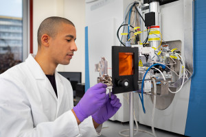
© ISAS / Hannes Woidich
Improving the performance of MALDI MSI sources
One requirement for the visualisation of small molecules in tissue sections is sufficient ion signal throughout the measurement, ideally without influences from matrix background or other analytes. Therefore, the scientists involved in this programme are dedicating a huge part of their work to improving the performance of MALDI MSI sources with regard to the overall ion signal, reduced ion suppression effects, increased coverage of lipids and metabolites in one MSI run. For this, the researchers are combining different ionisation sources with MALDI MSI, for instance ISAS’s flexible microtube plasma (FμTP). The scientist will then test the optimised ion sources for the analysis of lipids and metabolites in cells and cardiovascular disease-associated tissues (inflamed heart tissue after an infarction and Fabry Disease) obtained from mouse models and human samples.
Developing and optimising different sample preparation strategies
Another aspect that can significantly influence the quality of MSI results is sample preparation. Tissue washing steps may lower the total salt content, additives may improve ionisation efficiency, or chemical derivatisation can enhance the signal of select ompound classes and help elucidate the molecular structure of analytes. For this reason, the development and improvement of sample preparation strategies is part of the work in the MS-based imaging research programme. Specifically, these include
optimising protocols for MSI metabolites of the citric acid cycle,
minimising ion suppression effects by removing salts or suppression analytes,
on-tissue derivatisation methods to target low abundant or hard to ionise compounds focusing on steroids, oxidised lipids, sphingolipids,
chemical derivatisation methods to structurally characterise analytes or validate compound annotations, and
increasing the compatibility of MSI sample preparation with modalities such as fluorescence and Raman microscopy.
Library of quantified lipid and metabolite values
Quantification is difficult with MSI, because ion suppression effects could depend on the tissue type or the histological structures of the tissue. That is why the researchers at ISAS intend to combine their improved ion sources and optimised sample preparation strategies with absolute quantification results from established shotgun and liquid chromatography (LC) MS/MS experiments. One of their goals is to create a library of quantified lipid and metabolite data for a range of tissue samples using MALDI MSI and to compare the results with established shotgun and LC-MS/MS values. Another aim is to develop a software tool that allows to use these libraries tool to quantify compounds in preclinical samples (genetically altered mouse models) and clinical samples.
3D-printed funnel and ion-mobility spectrometer
Combination is also key when it comes to MSI set-ups. In the MS-Based Imaging research programme, the scientists aim to develop a miniaturised ion funnel and a 3D-printed stand-alone drift tube ion-mobility spectrometer that is compatible with MSI technologies to improve the ion transfer, its sensitivity, and enable separation of ion populations, respectively. It is expected that these devices can significantly improve the performance of MSI set-ups for molecularly resolved spatial profiling, are customisable in size thanks to 3D printing, and are therefore compatible with multiple MSI set-ups.