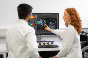
Modern imaging methods are regarded as a key technology in first-class medical research. At ISAS, the research programme 3D Molecular Pathology focusses on temporally and spatially high resolution visualisations and measurements of physiological and pathological states in whole organs, the tissue structures and cells of which they are composed of, down to the molecular components which are essential for the function of the cells.
Inflammation as a basis of many pathological processes & positive events
Various research groups at ISAS are working on different projects to elucidate the molecular and cellular processes that underlie immuno-vascular interactions under inflammatory conditions. The researchers investigate these cell-cell interactions, both in acute inflammatory processes and in chronic autoimmune disorders. Inflammation is the basis of many pathological processes in the human body. In addition to injuries or infections as triggers, also internal events like a vascular blockage can lead to an inflammatory reaction. Examples for these so-called sterile or aseptic inflammations (meaning no pathogens are involved in their development) are for example heart attacks, strokes, autoimmune diseases like rheumatoid arthritis, or cancer. Sterile inflammation is characterised by a massive infiltration of activated immune cells (“inflammatory cells") into the inflamed tissue and a systemic flooding (of the whole body) with soluble inflammatory mediators.
However, immune cells that migrate into inflammatory sites can also perform important positive tasks in sterile inflammation, such as the regeneration of tissue damage, the local restriction of inflammatory foci by encapsulation or the fight against tumours. For this reason, it is difficult to clearly classify the role of an immunological infiltrate as "harmful" or "beneficial". Both, the molecular context in which the immune reaction takes place and its timing in relation to the triggering event are essential when evaluating the impact of an immune response to sterile inflammation, and thus also for the question of how to treat patients most efficiently.

© ISAS / Hannes Woidich
Combination of complementary methods for full-scale analyses
Using Light Sheet Fluorescence Microscopy (LSFM), high-resolution Confocal Laser Scanning Microscopy (CLSM) and Raman Microscopy for example, scientists at ISAS identify and validate biomarkers to accelerate the early detection of adverse conditions such as cardiovascular or autoimmune diseases, and their impact on systemic integrity (for example resulting in an immune dysfunction). To translate this basic research into clinical practice, there is a close cooperation, for example, with the Institute for Experimental Immunology & Imaging at the University Hospital Essen. Moreover, the researchers develop complementary new microscopy techniques which are designed to massively increase the throughput of samples, and therefore the speed of analyses. In addition, the scientists use artificial intelligence (AI) to analyse entire organs down to the level of individual cells in experimental disease models in mice or in tissue and blood samples from patients. Depending on the microscope used, one individual sample can produce hundreds of images. Without AI, an in-depth rapid quantification and understanding of the biological information contained in these images would not be possible, nor would it be possible to administer it efficiently. Therefore microscopy is only one of many areas of application in medical imaging where AI is continuously revolutionising the processing of huge quantities of data.
The combination of LSFM and CLSM allows scientist to carry out a three-dimensional analysis of biological samples from the macroscopic to the subcellular level. However, in order to be able to characterise morphological and functional changes in inflammatory tissues with their fundamental mechanisms in molecular detail and over a period of time, scientists at ISAS combine time-resolved CLSM, LSFM and complementary analytical technologies such as mass spectrometry (MS), mass spectrometry imaging (MSI), and high-dimensional flow cytometry.
Striving towards a multimodal analytics workflow with non-destructive, integrative analyses
Since a disease mechanism is not only decisively influenced by the function of a biomolecule in a system but also its precise occurrence in time and space, combining microscopic methods with general and locally-resolved MS paves the way for entirely new diagnosis options in the future. At present, many of the stated imaging methods still inevitably lead to a destruction of the samples. This means that analyses are restricted to using individual techniques, which may also be mutually exclusive. This is problematic, especially regarding rare samples like human tissue biopsies, because comprehensive analyses are only possible to a limited extent. In the 3D Molecular Pathology programme, ISAS researchers therefore work on harmonising and combining complementary imaging and analytical methods with the aim of obtaining new non-destructive integrative measurement strategies. The purpose of this cross-scale multi-method concept – in the form of 4D analyses – is to enable the location- and time-resolved, quantitative in-vivo analysis of biologically relevant components at the cellular to molecular level. Key technical innovations are required to enable a truly comprehensive multimodal and multidimensional analysis, and therefore for an overall understanding of biomedically relevant processes. In the long run, these emerging new analytical technologies are supposed to be integrated into clinical diagnostics which in turn should lead to improved prevention and early disease diagnosis as well as personalised therapies.