Dortmund, 13th October 2025
When developing data-centric artificial intelligence (AI), the focus is on optimising training data. This approach is less common in biomedical research than model-centric AI applications. A team led by Dr Jianxu Chen from ISAS, with researchers from China, Germany and the USA, aims to change this and establish data-centric AI applications in bioimaging research. To this end, they have published a so-called framework (open source) in the journal npj imaging. The framework is intended to make it easier for scientists from the bioimaging community to apply AI in the analysis of image data.
Reliable AI systems are based on reliable data – that is the basic idea behind data-centric AI applications. Instead of constantly improving the model, researchers manually optimise the quality and representativeness of the training data with the support of AI applications. For example, they examine whether the data is clean, complete and balanced. This allows to continuously improve a defined AI model with refined measurement data. The counterpart to this is model-centric AI applications. Here, researchers use a fixed dataset to improve the AI model.

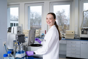
Dr Jianxu Chen heads the AMBIOM – Analysis of Microscopic BIOMedical Images junior research group.
© ISAS / Hannes Woidich
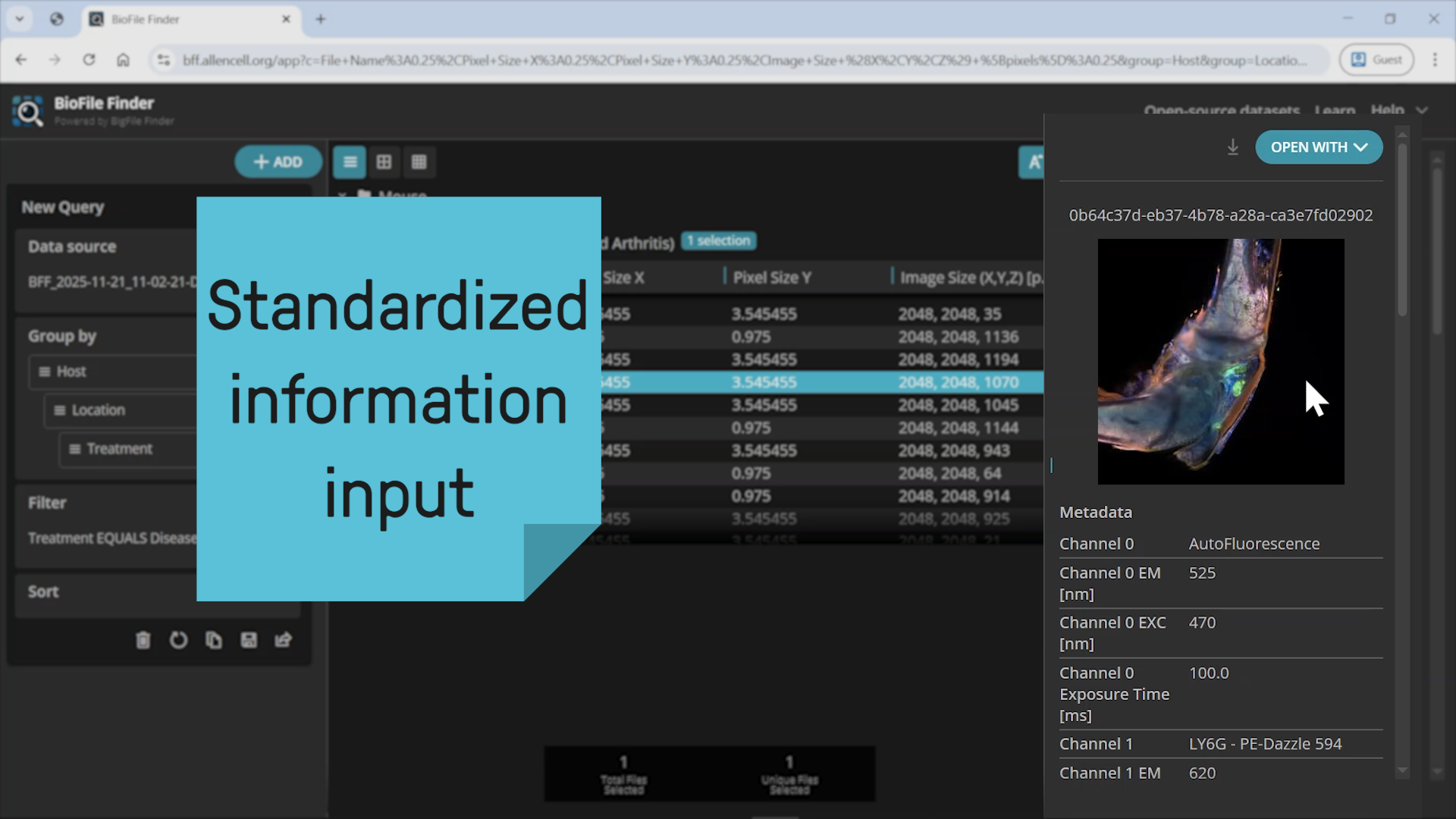
Dr Jianxu Chen, Head of the AMBIOM – Analysis of Microscopic BIOMedical Images junior research group at ISAS, is the corresponding author of the new publication. "Our framework is intended to serve as a common language for AI experts and biomedical researchers", explains the expert in biomedical image analysis and deep learning. A framework describes a collection of programming tools and libraries that help developers to create and train AI models more easily. In this case, it provides an organized overview of all relevant AI methods and techniques in the context of bioimaging that help biomedical scientists think through their bioimaging problems in a data-centric mindset.
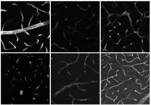
Examples showing the diversity of a vasculature dataset from a real biomedical study, with varying morphology, intensity, signal-to-noise ratio, etc. The “BioData-Centric AI” framework provides a systematic approach for biomedical researchers to tackle such real-life challenges.
© J. Wanzel, J. Lampe & M. Schwaninger: Institute for Experimental and Clinical Pharmacology and Toxicology, Center of Brain, Behavior and Metabolism (CBBM), University of Lübeck, Germany; DZHK (Deutsches Zentrum für Herz-Kreislauf-Forschung e. V., German Research Centre for Cardiovascular Research), Hamburg-Lübeck-Kiel, Germany.
Chen demonstrates the advantages of the new four-stage framework using an example that he developed with AI researchers worldwide and scientists from the field of biomedical research. For this purpose, he and his co-authors collected over 800 three-dimensional microscope images for the segmentation of a vascular structure. Rather than evaluating this large amount of data manually, as has been the norm for model-centric approaches to date, the researchers used the raw data to pre-train an AI model. Using this initial AI application, they identified the most representative images from the dataset to train an AI model for further evaluation. They then continuously improved this model by, for example, manually analysing microscope images that the model had difficulty with, using these to further enhance the model.
Framework builds bridge between AI development and biomedical research
In the cited example, the researchers used basic algorithms that are accessible via GitHub (editorial note: We would like to inform you that after activating the link, data will be transmitted to GitHub.). Integrating further complex algorithms is also a possibility for the future. Additionally, the framework could be applied to other research questions in biomedicine. The authors emphasise that model-centric and data-centric approaches are by no means mutually exclusive and could be combined in future research.
The framework is intended to serve as a guide for two target groups in particular: scientists in biomedicine and developers of AI methods for biomedical research. "The framework helps biomedical scientists integrate AI applications into their research in a meaningful way, without requiring them to know everything about AI", explains Chen. For developers, the paper provides an overview of the possibilities of data-centric AI and guidance for future developments.
Publication
Cao, J., Wenzel, J., Zhang, S., Lampe, J., Wang, H., Yao, J., Zhang, Z., Zhao, S., Zhou, Y., Chen, C., Schwaninger, M., Yang, J., Chen, D. Z., Chen, J.
(2025) Rethinking deep learning in bioimaging through a data centric lens. npj imaging 3, 29.
(Anna Becker)