Skip to content

Where the Human Brain Reaches its Limit

Dortmund, 24th May 2022

Microscopy is just one of many fields of application in medical imaging that can hardly be imagined without artificial intelligence (AI) in the processing of enormous amounts of data. Because humans cognitively reach their limits despite existing programmes for image evaluation, the AI experts at ISAS are developing eyes and brains for computers. Prof. Dr. Anika Grüneboom, head of Bioimaging, and Dr. Jianxu Chen, head of the AMBIOM junior research group, each report in their statements why the interdisciplinary cooperation of their research groups is vital to their work and what the advantages are.

Prof. Dr. Anika Grüneboom und Dr. Jianxu Chen sprechen über Aufnahmen von Blutgefäßen im Unterkiefer der Maus.

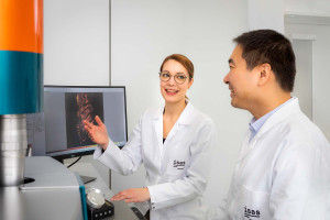
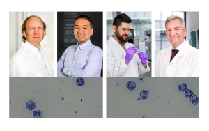
Prof. Dr. Anika Grüneboom (left) and Dr. Jianxu Chen talk about images of blood vessels in the murine mandible (lower jaw of the mouse), which were previously taken using the light sheet fluorescence microscope.

© ISAS

Prof. Dr. Anika Grüneboom, head of Bioimaging

“The cells in our blood vessels talk to each other. How they interact with each other, how far apart they are – all this provides us with important insights into inflammatory diseases. The cells of our immune system communicate with each other as well as with the endothelial cells that line the innermost layer of the blood vessels. This communication controls where immune cells attach to blood vessels, migrate through them, and then enter surrounding tissues to fight inflammation. In autoimmune diseases such as rheumatoid arthritis, however, the infiltration of immune cells into tissue does not lead to healing of the inflammation, but rather worsens it and ultimately causes the disease to become chronic. Therefore, we would like to identify criteria that will allow us, in the future, to detect these processes in the blood or tissue of each individual person before the actual onset of disease – that is, long before these inflammations become chronic.

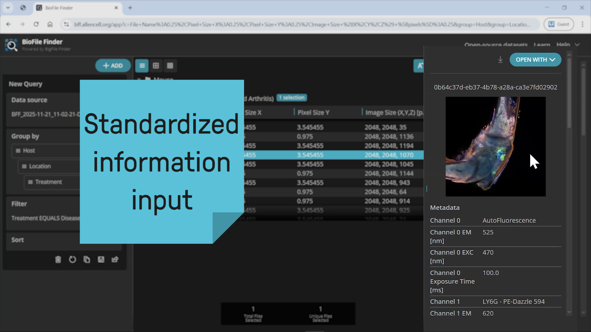
For example, in my research group we examine the inner layer of blood vessels under the light sheet fluorescence microscope. We are studying which cells are involved in inflammatory processes, what the cell walls look like, how far apart the cells are from each other and how the cells communicate with each other. The light sheet fluorescence microscope expands the laser beam like a sheet of paper, and this thin disk of light that is created illuminates each individual plane of our samples and takes a picture of each plane. We later assemble the individual images into a 3D model on the computer. On average, we can produce well over 500 images from a single sample under the microscope, and we usually work with more than 20 samples per test series. The many images that the microscope creates pose a great challenge for us. Although there are computer programs available today, we researchers still spend a lot of time evaluating all the information from these images. Artificial intelligence can help us with this task much better than an individual computer program."

What does AI mean for the analysis of microscopic images?

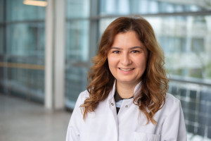
Dr. Jianxu Chen, head of AMBIOM

“Communication is key – not only between the cells in our body, but also between our research groups. With Artificial intelligence (AI), we can merge high amounts of data. There a mainly three important things that machines can do that humans simply cannot. That is why there is a high potential for a deeper synergy between microscopic image analyses and AI.

First of all, AI lets us analyse way more images and thus gives us a higher throughput than an ordinary computer programmes. We can have intelligent machines analyses large amount of pictures with a precise outcome. Second, a big advantage is that AI can see things that are not visible for the human eye. AI is able to interpret every information in the images. For example: The human eye can look at the images and concentrate on the thickness of the blood vessels. AI can look at the same images, but evaluate more: not only the blood vessels’ thickness, but also how rough their surface is, what is happening next to their walls, and even more. Third, our human brain is limited, whereas intelligent machines do not have that limitation. They are able to extract information and build knowledge that the human brain can hardly process. That is why my research group programs algorithms that serve as the eyes and brains for computers.

When we design our algorithms, we make sure that we are accurate and at the same time sustainable in terms of using power resources. What is also crucial is that we believe science should be transparent. Therefore, our analyses are fully reproducible, because my team’s work is based on open source. It is important to us at ISAS that scientist all around the world have access to the AI methods that we develop here in Dortmund." 

The MSCoreSys associated junior research group AMBiOM – Analysis of Microscopic BiOMedical Images is funded by the Federal Ministry of Education and Research (Bundesministerium für Bildung und Forschung, BMBF) under the funding reference 161L0272.

Share

Further articles

28th March 2024

New “Green” Microscopy: Less Electricity, but More Information on Immune Cells in Return

Advanced technologies, such as high-resolution microscopes, produce large amounts of data. And these again consume large amounts of electricity. In addition, there are refrigerators for samples, fume cupboards and small technical devices. While ISAS is upgrading to become greener, researchers at the institute are already working on methods to make microscopy more energy-efficient in general.

Das Bild zeigt eine schematische Darstellung der Datenverarbeitung in der Mikroskopie.
13th March 2024

Cirrhosis of the Liver: Migrating Immune Cells Act as an Early Warning System

The life expectancy of patients with liver cirrhosis depends crucially on the occurrence of disease-associated complications, such as infections. Until now, however, an ability to predict these at an early stage has been lacking. A problem, often hindering doctors from administering antibiotics or even performing a liver transplant in time. Researchers at ISAS led by Prof Dr Matthias Gunzer therefore investigated the question: Could the mobility of certain immune cells be the decisive indicator of an impending deterioration in health?

27th February 2024

3 Questions for… Dr Christopher Nelke

As a participant in the Clinician Scientist programme and physician at the Clinic for Neurology at Düsseldorf University Hospital (Universitätsklinikum Düsseldorf, UKD), Dr Christopher Nelke researches neuromuscular diseases. In this interview, he talks about his two-week guest stay at ISAS and the challenges that arise between the hospital bed and research.

Das Bild zeigt Dr. Christopher Nelke im Labor. In den Händen hält er eine Probe. The picture shows Dr Christopher Nelke in the laboratory. He is holding a sample in his hands.
20th February 2024

SARS-CoV-2: The Very Latest Methods Clarify the Active Agents and the Mechanism of Action of Ancient Self-Medications

Prophylactic, soothing or even healing agents, mostly natural substances, have been known to natural medicine since ancient times. But what about viral infections? Could infusions made from sage or perilla also be used against SARS-CoV-2 infections -as a prevention or an aid to healing? An interdisciplinary team of researchers led by Prof Dr Mirko Trilling from the Faculty of Medicine at the University of Duisburg-Essen (UDE) and scientists at ISAS investigated these questions during the coronavirus pandemic.

Das Bild zeigt Prof. Dr. Mirko Trilling mit verschränkten Armen, an einer Wand lehnend. The picture shows Prof Dr Mirko Trilling with his arms folded, leaning against a wall.
7th February 2024

A Long Sought-After Combination Method in Mass Spectrometry

Researchers who analyse complex samples using mass spectrometers are often faced with the challenge that the substances they contain are fundamentally different. Some are chemically polar, others are non-polar. Until now, this has required two complex separate analyses. But a researcher at ISAS has developed a method with which even less polar substances can be included in a standard mass spectrometric analysis for polar biological substances.

Daniel Foest steht im Labor und hält ein Papier mit einer Leberprobe, die er am Massenspektrometer untersucht.
12th January 2024

“My research is literally hard work"

Darleen Hüser is looking for the immuno-cellular fingerprint of rheumatoid arthritis. In this interview, the doctoral student reveals her razor-sharp research and why she needs different microscopes.

Das Porträt zeigt ISAS-Doktorandin Darleen Hüser aus der Arbeitsgruppe Bioimaging.
21st December 2023

Science Slam: Humorous Science Communication Is Fun for Everyone

Talking lab equipment, artificial intelligence and expertise from the North Pole - this colourful mix of topics characterised the latest Science Slam at the institute. Four ISAS employees demonstrated with their specialist knowledge and plenty of humour how science communication can be fun for everyone involved.

Luisa Becher fotografiert die vier Teilnehmenden des ISAS Science Slam.
20th December 2023

The Art of Balancing: Accuracy in Image Analysis

What challenges in analysing microscopic images can be mastered with artificial intelligence if the latter is incorporated at an early stage? Why should researchers think about the target metrics of image analysis when planning their experiment? Dr Jianxu Chen has now put together his tips, recently published in the journal Nature Methods, as a kind of checklist for researchers.

Die Abbildung zeigt eine Wage und symbolisiert das Gleichgewicht zwischen Analyse und Genauigkeit bei der Validierung von biomedizinischen Aufnahmen.
1st December 2023

Bone research: ISAS participates in the new "DIONE" Collaborative Research Centre

The nationwide project recently funded by the German Research Foundation is focussing on inflammation-induced bone loss. Researchers from Dortmund, Dresden, Erlangen/Nuremberg and Ulm want to find out exactly how inflammatory diseases - such as rheumatoid arthritis or intestinal diseases - damage bones. Among other things, their research aims to help identify new therapies for skeletal-associated diseases.

Bone structur with osteoporosis.