Dortmund, 10th August 2022
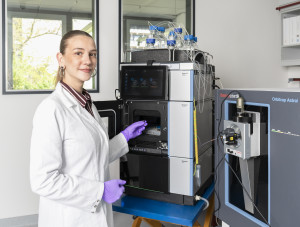
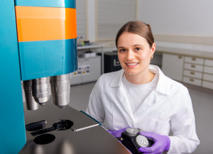
Neutrophil granulocytes – in short neutrophils – are a type of white blood cells (leukocytes), and can promote tumour growth although their task as the body’s police is the exact opposite. To understand why neutrophils cannot only be tumour-inhibiting but also tumour-promoting, Susmita Ghosh (Bio-Fluorescence) wants to unravel the molecular makeup of these immune cells in tumours. However, the 25-year-old has only few samples from mice available for the analysis with a mass spectrometer. In order to avoid mistakes that could damage the biopsy material, a robot could assist Ghosh in the future. Therefore, Julia Rauch, a PhD student in the Proteomics research group, helps with its technical implementation. Together, the junior researchers are working on enabling the robot to complete the entire sample analysis – from pipetting to peptide elution – in a precise and fast manner.
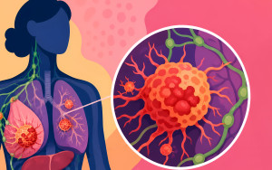
As immune cells of the innate, non-specific immune response, neutrophils are essential for the human organism. For example, they repel infectious agents or fight inflammation. In case of a tumour, the body’s blood stem cells in the bone marrow react, for example, by producing more neutrophils. The fact that these immune cells move fast but react in a non-specific way, lets them play an important role in carcinogenesis – the process that enables the development and growth of tumours. Recruited by signalling proteins of the cancer cells, neutrophils enter the tumour microenvironment. Due to molecular processes, these tumour-associated neutrophils (TANs) can have tumour-promoting or tumour-inhibiting effects in said microenvironment. The former are especially dangerous for cancer patients. They promote tumour growth, encourage metastases and worsen the prospect of a successful chemotherapy.
The molecular differences between these two types of TANs are still unknown. Therefore, Ghosh is examining the type and amount of proteins in the neutrophils for her doctoral thesis. Her goal is to identify the proteins which are typical for TANs. In this way, the biologist, who has been working as a PhD student at ISAS since October 2021, eventually wants to identify possible targets for new drugs, for example against skin cancer.
Melanoma samples do not forgive mistakes
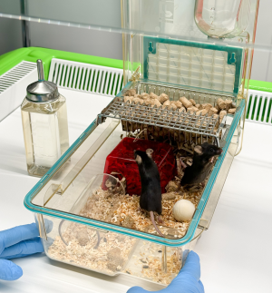
The 25-year-old is mainly researching skin cancer by using melanoma samples from mice. However, she finds the biopsy material challenging: “The amount of neutrophils in the mice tumours is quite small. In addition, the number of proteins in the neutrophils is also low,” Ghosh explains. This is why she has to work carefully and precisely while preparing the samples for the mass spectrometer. After all, the junior researcher not only has to extract the proteins from the neutrophils, but she also has to clean them by dividing the peptides (chains with less than 100 amino acids) from the complete proteins (chains with more than 100 amino acids). The spectra of the peptides in the mass spectrometer later convey which and how many proteins are present in the neutrophils. In order to, figuratively speaking, cut the neutrophils’ proteins into peptides the Indian native uses a manual method. However, this procedure takes time and is sometimes imprecise. The consequence: the samples become unusable.
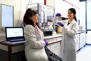
Susmita Ghosh showing Julia Sophie Rauch the magnetic beads which help remove contaminants from the protein solution.
© ISAS
Robot is supposed to enable an automated proteomics workflow
When Rauch, who has already worked with various robots in the lab, heard about Ghosh’s challenge, she instantly suggested to apply the so-called SP3 protocol: the Bravo robot could take over all steps from pipetting, digestion, to the elution of proteins and peptides with the help of magnetic beads. The machine has several advantages: “A human can process about ten samples of this sensitive biopsy material per day. The Bravo robot manages 96 samples with less or hardly any mistakes in one run”, says Rauch who has been conducting research at ISAS for three years.
It is a helpful coincidence that she and Ghosh share an office: what started as a spontaneous idea in a chat between two colleagues a few months ago, has grown into a collaboration between two research groups. Since February 2022, the two junior researchers have been working on transferring the existing manual workflow to the robot. They are currently trying to ensure error-free pipetting results. Even though the volumes differ after pipetting and supply shortages hinder them from getting supplies for the robot, Rauch and Ghosh stay optimistic. Because if they succeed, other researches will also benefit from their efforts.
Benefits for cancer research & scientists
In the future, the Bravo robot adjusted by Rauch and Ghosh could be of help when it comes to other challenging samples. “An automated, error-free workflow could also optimise the analysis of other leucocytes, for example lymphocytes,” Ghosh explains. For Rauch, the overall advantages go hand in hand: “Frequent manual pipetting puts a strain on the tendons in hands and arms. I know many colleagues who suffer from tenosynovitis because of that. Therefore, the robot could ease the daily lab routine in every respect”.
Bravo robot: All-rounder in the lab?
Rauch and Ghosh’s goal is to make the robot fit for using the SP3 workflow automatically including the following steps: After having extracted the proteins from the neutrophils with a chemical solution (lysis buffer) during sample preparation, the robot is supposed to add tiny magnetic beads to the protein solution. These bind to the proteins. In the next step, the machine will place the 96-well plates – microplates with space for 96 sample containers – on magnetic disks. The magnetic field will concentrate the beads-protein-mixture on one spot. This step helps with the removal of unwanted contaminants, for example remains of the lysis buffer. After that, the enzymatic digestion will split the proteins into peptides. Since the eluted peptides do not bind to the magnetic beads, they can be extracted for the analysis with a mass spectrometer.
(Bettina Dirauf)