Dortmund, 15th July 2022
Knee prostheses, hip implants or cardiac pacemakers – due to medical progress, there are numerous implants designed to support the human body. However, despite all sterile preparations, implants also carry a risk of infections. To prevent these, Kaja Reiffert, PhD student in the Bioimaging research group, is developing a strategy using silver.
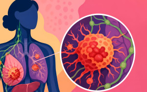
When bacteria grow on the implant’s surface – most commonly during its insertion in surgery – an infection likely develops. The bacteria build their own environment and thus create the perfect conditions to grow and multiply. A biofilm, a community of bacteria, develops. Once a biofilm has formed, it is very solid and difficult to combat. Antibiotics resistance makes the treatment even more difficult. Inflamed tissue around the implant causes pain and, in the worst case, there is a risk of blood poisoning. The consequence: In case of an infection, the implant has to be removed.
Small particles, big effect
Reiffert is working on a strategy using silver in order to prevent infections from occurring in the first place. Silver ions have an antimicrobial effect. On the implant’s surface, they could protect it from bacterial colonisation. To achieve this antibacterial effect as efficiently as possible, Reiffert uses ultra-small silver particles. When they are exposed to an aqueous environment, they release antimicrobial silver ions. The size of the particles is of great importance, because during contact the following applies: The smaller the particles, the bigger is their surface and thus the greater the rate of release of silver ions. “Imagine a cube. If you divide a big cube into eight small ones, you have a much bigger surface area in total. This increase in surface area can ensure that more ions are released,” the 24-year-old explains. The junior scientist works with ultra-small particles that are just one to two nanometres in size. With these, she hopes to increase the antimicrobial effect on the implant.
Silver, gold & platinum
The effect of silver particles that are the size of one to two nanometres on the human body is yet unknown. “The first question is whether toxicity can occur in the body; in other words, whether the silver particles are toxic for humans. It is not enough to study only the effect on the bacteria. We also have to observe whether the silver ions affect human cells and how,” the biologist points out. She analyses the effects on so-called mesenchymal stem cells, cells of the supporting and connective tissue.
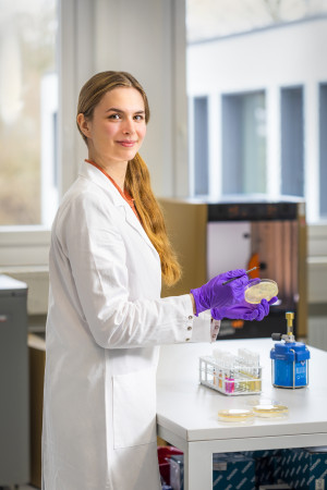
For her dissertation, Kaja Reiffert examines how ultra-small silver particles can prevent infections in implants, for example with bacteria like Escherischia coli (E. coli).
© ISAS
The special thing about these stem cells is that they can develop into different types of cells. In the human body, they can become bone-, cartilage- or fat cells. Researchers refer to this development as stem cell differentiation. Platinum has an osteogenic promotion effect; that means it promotes the differentiation of bones cells. This effect could strengthen the connection between bone and implant and thus prevent inflammation. That is why the young researcher also wants to study the effect of silver in combination with platinum in her dissertation.
The same is true for the combination of silver and gold. Promotive effects on ostegenic differentiation have already been observed: “Gold nanoparticles are already being intensively studied for biomedical research. That is why we know more about their chemical and physical characteristics than those of silver and platinum.
However, the nanoparticles used in these studies are much bigger than the ones we use. The exact effects of ultra-small gold nanoparticles are actually still unknown. As with silver and platinum, we expect an increased ion release and thus a more efficient effect. We want to use this to optimise the osteogenic promotion effect.”
What are nanoparticles?
Nanoparticles are particles smaller than 100 nanometres (nm). One nanometre corresponds to one millionth of a millimetre. That is about 1000 times smaller than the diameter of a human hair. In the case of ultra-small nanoparticles, scientists usually speak of a size between 1-3 nm.
Combination of analytical methods required
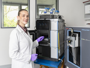
During the first steps of her research, Reiffert wants to find answers to the following questions: How do the silver particles get into the cell? And what happens once the cells have taken them in? For her analysis, Reiffert uses various imaging techniques. “With the help of the confocal microscope, I want to find out whether nanoparticles can penetrate the cells and where they are localised within the cell. For this, I make different cell compartments visible with fluorescent dyes or antibodies as a marker and use fluorescence-marked nanoparticles,” the PhD student explains. Since it is not yet clear whether the nanoparticles’ fluorescence marker influences the images or the localisation, Reiffert also uses a transmission electron microscope. Transmission electron microscopy does not require fluorescent dyes. Contrasting makes definable cell structures, the organelles, visible. With the transmission electron microscope, Reiffert takes high-resolution images, which are necessary for the depiction of ultra-small nanoparticles. Later, Reiffert also wants use a flow cytometer to characterise the cell populations and determine how efficiently the cells take in the nanoparticles and how the ultra-small particles affect the cells’ vitality and differentiation. The researcher sums up: “Only the combination of different analytical methods yields all the information we need to study the nanoparticles’ effect on bacteria and human cells and evaluate their suitability for implants comprehensively.”
About the DFG project
Kaja Reiffert is doing her PhD at the University of Duisburg-Essen. The title of her dissertation is "Ultra-small Mono- and Bi-metallic Nanoparticles as a Possible Prevention Strategy of Implant-associated Infections" (Ultrakleine mono- und bimetallische Nanopartikel als mögliche Präventionsstrategie Implantat-assoziierter Infektionen). Her work contributes to the research project "Synthesis, structure, and biological effects of ultrasmall (1-2 nm) bimetallic silver-platinum nanoparticles". The German Research Association (DFG) funds the project under the number 452179459.
(Nadine Gode)