Dortmund, 2nd February 2022
It has been five years since Mohammad Ibrahim Alwahsh came to Germany as an exchange student. Back then, he was doing his Master's in pharmaceutical science at Al Zaytoonah University of Jordan. Impressed by the quality of the research in Germany, he decided to stay for his doctorate. Since 2018, the 27-year-old has been working on methods of analytical toxicology to improve the treatment of a rare form of cancer in the thymus gland. With success – Alwahsh and his colleagues at ISAS and Heidelberg University recently succeeded in using their method to measure the live reaction to chemotherapeutic agents in 3D models for the first time. Moreover, they were able to highlight possible therapeutic options for patients with thymoma and thymic carcinoma.
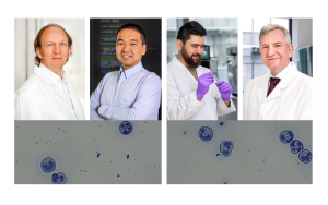
Thymoma and thymic carcinoma are both tumours in the thymus gland, a central organ of the lymphatic system and thus of the immune defence system in humans. Both are so-called Thymic Epithelial Tumours (TETs). According to information published by the American Society of Clinical Oncology (ASCO) in 2021, about 400 people in the US are diagnosed with thymoma every year. Accordingly, Thymic carcinoma is even more rare and makes up about 20 percent of thymic tumours. Rare diseases like these often present researchers with a huge problem: "Thymoma and thymic carcinoma are so rare that it is hard to find human tissue samples," Alwahsh explains. That is why he cultivates the cancer cells in the laboratory.
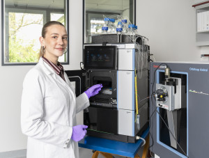
The 27-year-old reproduces the tumours as spherical 3D structures, so-called spheroids or organoids. "The tricky thing about cancer therapy is the physiologically different conditions deep inside a tumour, which cause tumour cells to behave differently there than they do on its surface", he points out. 3D models can imitate this process – and are thus more realistic and persistent in terms of reaction to medication than common two-dimensional models. According to Alwahsh, it is therefore better to test new therapies for TETs in the laboratory (in vitro) not only on 2D but also on 3D models before moving on to animals or humans (in vivo). The pharmacist uses a nuclear magnetic resonance (NMR) spectrometer he developed and patented at ISAS while working in the Bioresponsive Materials group during his doctorate.
Hourly updates on a drug’s effect thanks to NMR spectroscopy
Until now, there has been a lack of methods to conduct a live analysis of 3D models to see how they react to drugs such as chemotherapeutic agents. Alwahsh's analytical technique enables him to observe the metabolites of a living 3D tumour model over a long period of time after applying the medication: He can inject chemotherapeutic drugs into the spheroids via a probe and observe live how the cells react. Over two days, the NMR spectrometer measures the metabolic activity of the cells every hour so that the researcher can see how the drug affects them. Particularly exciting and new: The scientist can link the metabolites to specific areas within the tumour. This allows him to adjust the medication accordingly to ensure that it not only attacks the outer cells of the tumour, but also the persistent, possibly dormant ones inside.
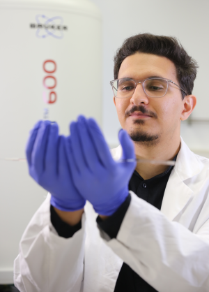
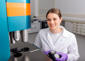
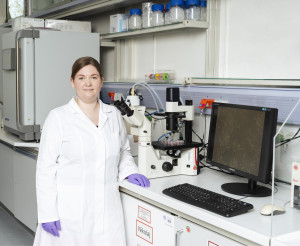
The spheroid/organoid in the glass incubator is so tiny that Alwahsh can barely see it with his eye.
© ISAS
Context: Research programme Biomarker
The work that Alwahsh and his colleagues carried out at ISAS was part of the Biomarkers research programme. The aim of all the work in this research programme is to identify biomarkers for early diagnosis or personalised therapy using a combination of different analytic techniques. Reliable markers expand the possibilities of evidence-based diagnostics in modern medicine, allowing for differentiated and individualised therapy.
"One of the strengths of our method is that the spheroids remain undestroyed"
Alwahsh's project is an interdisciplinary one, which was only possible through cross-group collaboration in the first place, he emphasises: "Teamwork was really the key to success in this project. Besides me as a pharmacist, the project involved physicians, engineers, chemists and physicists.” To ensure that the tumour can survive the two-day measurements and stay viable, the researchers had to come up with a complex solution. Outside the NMR spectrometer, they placed an incubator-like device that guarantees the optimal temperature and oxygen saturation for the growth of the tumour cells during the analyses. A microchip supplies the tumour with nutrients and the drug to be tested. In this way, the scientists recreate the situation in the human body in the best possible way. "One of the strengths of our method is that the spheroids remain undestroyed during the measurement," Alwahsh adds. After the analyses, the 3D models can continue to grow in an incubator outside the NMR spectrometer until the researchers perform new tests.
Including optical parameters in future analyses
At the end of 2021, shortly after his son was born, Alwahsh also celebrated the submission of his dissertation. However, that is no reason for the young father to rest on his laurels. He is currently working on validating the obtained therapeutic options. Alwahsh also wants to establish a new technique that integrates imaging techniques such as fluorescence microscopy into his method. This way, he would like to include optical parameters in his future analyses. A scholarship from Al Zaytoonah University of Jordan for his dissertation brought Alwahsh to ISAS and him to realise: Wherever his future path will take him, even far from home, far from his relatives and friends, he can flourish as a scientist.
(Cheyenne Peters)