Skip to content

Guest Presentation: New Framework for Stem Cell Analysis

Dortmund, 27th January 2023

Understanding and predicting the behaviour of cells in normal, regenerative and pathological contexts – these are the goals of the Allen Institute for Cell Science. To achieve this, the non-profit research organisation develops multi-scale visual models of cell organisation, dynamics and activity. Its software programmes are freely accessible to researchers worldwide (open source). They are part of the Allen Cell Explorer, a portal for cells, cell biology, data and tools. The portal provides insight into the organisational diversity of human stem cells and delivers matching open data, cell lines, plasmids and models. Human stem cells from the test tube, so-called induced pluripotent stem cells, are the subject of a recently published paper in Nature.

How does a subset of expressed genes dictate cellular phenotype – the visible properties of cells? Finding the answer to this question is a considerable challenge for scientists due to the large number of molecules involved, their combinatorics and the abundance of cellular behaviors that determine this phenotype. Scientists at the Allen Institute for Cell Science have recently succeeded in developing a framework for the analysis of integrated intracellular organization and its variations in human iPS cells, based on datasets of 200,000 living cells in 3D and a set of 25 key structures.

 A framework is a basic software scaffold that developers can use to build computer applications. The framework developed at the Allen Institute for Cell Science allows for programming analysis software that converts raw image data of iPS cells and their structures into dimensionally reduced, quantitative analyses.

Portrait Susanne Rafelski.

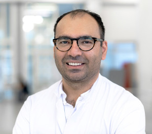
Dr Susanne Rafelksi is a biochemist and has been Deputy Director of Scientific Programmes at the Allen Institute for Cell Science since 2020.

How the framework will exactly advance stem cell analysis is the topic of Dr. Susanne Rafelski's presentation, "Integrated intracellular organization and its variations in human iPS cells," at ISAS on February 7.

Tuesday, February 7
10.30 am
Webex

https://t1p.de/rk032
Meeting ID: 2730 175 0712
Password: uMegm3Dw5B5

Rafelski is deputy director of scientific programmes at the Seattle institute – and corresponding author of the Nature publication. ISAS has a cooperation with the Allen Institute for Cell Science. The AMBIOM working group led by Dr Jianxu Chen is in charge here. The 34-year-old worked at the Allen Institute for Cell Science for four years before joining ISAS.

(Sara Rebein)
Share

Further articles

Endogenous Antioxidant Promotes Breast Cancer Cells in the Lung

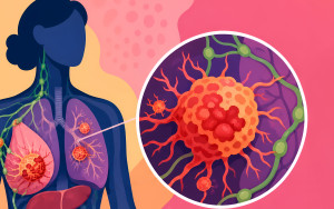
Oxidative stress puts pressure on metastasising tumour cells – yet they can activate defence mechanisms to cope with it. Prof. Dr Dr Alpaslan Tasdogan explains the specific challenges faced by breast cancer cells in the lungs. In the interview, he discusses the therapeutic potential of specifically targeting these defence mechanisms of tumour cells in the future.

Prof. Dr. Dr. Apaslan Tasdogan.

An AI Agent That Understands the Language of Science

A colleague who never sleeps, never gets tired and completes complex analyses in minutes: a new AI agent for biomedical research is to be developed at ISAS.

Endogenous Antioxidant Promotes Breast Cancer Cells in Lungs

How do breast cancer cells manage to establish themselves in the lungs and survive there? Researchers at ISAS are investigating which metabolic processes help the tumour cells to adapt to their new environment. The findings provide new insights into the early stages of metastases.

From Human Stem Cells to Mice: Research into New Treatments for Heart Failure

What can human stem cells tell us, and why are mice still essential for developing new therapies for people with heart failure? In our collaborative project HI-FIVE, scientists and physicians work with both animal-free methods and, for some research questions, with mice. In this article marking World Day for Laboratory Animals, our animal welfare officer and the lead researcher of HI-FIVE explain why.

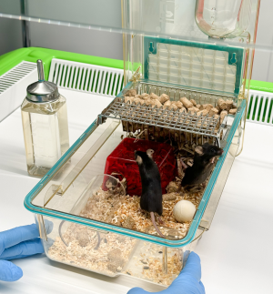
Mäuse in einer Haltungseinheit mit rotem Häuschen, Nistmaterial, Holzkugel und transparenter Röhre zur Beschäftigung und als Rückzugsmöglichkeit.

Four Perspectives on a Shared Discovery

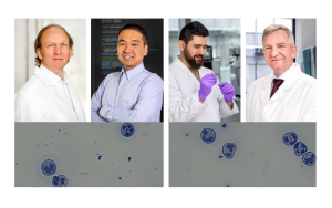
Although CD177⁺ and CD177⁻ neutrophils look identical under the microscope, they are in fact not the same. Four of the authors of “CD177 Deficiency Defines a Stable Subtype of Human Neutrophil Granulocytes with Tumour-Promoting Activity” (Advanced Science) explain how they identified the functional differences between these immune cells and which combined analytical methods made their discovery possible in the first place. This insight into their research demonstrates how interdisciplinary collaboration can reveal previously undiscovered information.

Das Foto zeigt im oberen Teil Porträts von vier Wissenschaftlern. Von links nach rechts: Prof. Dr. Matthias Gunzer, Prof. Dr. Jianxu Chen, Dr. Belal Alshaar, Prof. Dr. Albert Sickmann. Im Teil unter den Porträts sind Mikroskopaufnahmen von CD177-definierten Neutrophilen zu sehen.

3 Questions for... Neele Rottmann

In the lab, Neele Rottmann prepares human and animal samples for mass spectrometry analysis. As part of the HI-FIVE project, she is helping to identify changes at the protein level in heart failure. Why does she spend so much time in the lab working on separations? The technical assistant shares the answer in this interview.

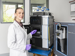
Neele Rottmann is wearing a white lab coat and purple gloves and is operating a mass spectrometry analyser in a laboratory. Next to her is a liquid chromatography system connected to a mass spectrometer via several tubes. There are numerous sample vials on the instrument. As she places a sample into the system, she looks into the camera.

High-tech against Cardiovascular Diseases: 3D Images of Blood Vessels

How do atherosclerotic plaques form in the coronary arteries? At ISAS, researchers are investigating cellular changes in human coronary arteries using techniques such as 3D imaging. During her internship, early-career researcher Leonie Menzel used a light sheet fluorescence microscope to analyse the spatial distribution of individual cells within the tissue. She thereby gained valuable methodological experience for her Master’s thesis.

3 Questions for Lukas Fu

How can AI support the analysis of biomedical images? This was the question Lukas Fu explored during his school internship at ISAS. The 15-year-old also successfully completed his first programming project.

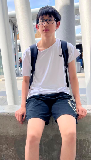
Lukas Fu, a 15-year-old with short black hair. He is wearing glasses and is sitting on a low wall.

Separating Fluorescence Signals Faster and More Precisely with AI

When different fluorescence signals overlap during microscopy, their clear assignment to specific biological structures becomes difficult. To address this common problem, Dr Davide Panzeri is developing AI-based signal separation methods. The biophysicist has been awarded a Marie Skłodowska-Curie Fellowship by the European Union for his promising research project.

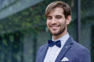
Porträt Dr. Davide Panzeri.