Dortmund, 21st November 2023
Fabry disease is a treacherous illness that gradually destroys the heart and other organs and is often not detected until it is too late to intervene. In future, a new diagnostic method tested at ISAS may help to diagnose thousands of sufferers in Germany alone at an earlier stage. Ultimately, other patients with cardiovascular diseases – the most common cause of death worldwide – might also benefit from the progress being made in the area.
The approach is based on Raman spectroscopy, one of the more recent spectroscopic imaging methods used in biomedicine. Biomedical spectroscopy – including the well-known nuclear magnetic resonance spectroscopy, which is based on measuring the magnetic momentum of nuclei – is becoming increasingly better at identifying illnesses using specific biomarkers, says Prof Dr Kristina Lorenz, head of the Translational Research department and the Cardiovascular Pharmacology research group.
“We deployed a special version of Raman spectroscopy, called coherent anti-Stokes Raman spectroscopy or CARS (see info box), to see if we can detect changes in the heart at early stages of the disease, changes that are caused by Fabry disease,” explains physicist Dr Elen Tolstik, who has been conducting research in the Cardiovascular Pharmacology research group since 2018.

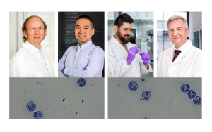
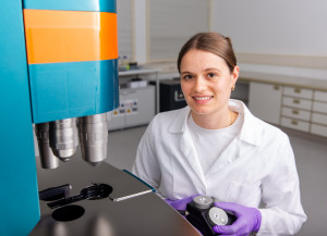
Prof Dr Kristina Lorenz is a pharmacologist; at ISAS she heads the Translational Research department and the Cardiovascular Pharmacology research group.
© ISAS / Hannes Woidich
RAMAN-SPEKTROSKOPIE & CARS
Raman spectroscopy uses the phenomenon of Raman scattering, where the light from molecules is scattered inelastically while their wavelength fluctuates. With the help of high-precision measurements, it is possible to obtain specific information from the process on the properties of the light-scattering molecules, for example, their chemical composition, structures and molecular dynamics. In contrast to simple Raman spectroscopy, with CARS two intense laser beams with different wavelengths selectively excite certain molecular vibrations.The resulting coherent scattering uses a significantly higher signal to penetrate deeper into the layers of tissue such that large structures can be analysed more quickly, says Tolstik.
Fabry disease – varied symptoms make diagnosis more difficult
The trigger for Fabry disease is a gene defect that causes too little or none of an enzyme named alpha-galactosidase A, or α-GAL A, to be produced in the body. In healthy people, the enzyme is present in virtually all cells of the body, where it breaks down what are known as glycosphingolipids – fats that are involved in creating cell membranes. Without α-GAL A and the breaking down of lipids it initiates, these lipids accumulate – they are “stored” as it were – which damages the cells over time. For this reason, Fabry disease is also called a “storage disorder”. Alongside organs such as the heart and the kidneys, the blood vessels and nerves are often affected.
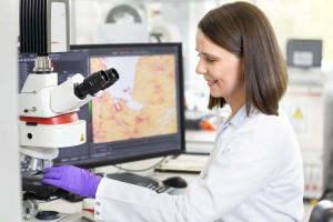
If treatment is commenced at an early stage, patients have good chances of survival, but in most cases the disease is diagnosed too late.
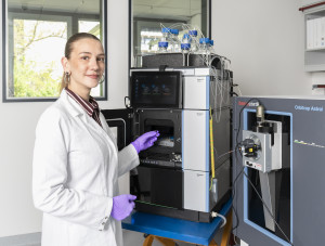
Dr Elen Tolstik
The damage to tissue caused by accumulations of lipids becomes apparent during the teenage years in some cases due to severe joint pains and changes in the skin. Typical signs are, for example, reddish-purple lumps and discolourations on the lower body. But this metabolic disorder may also cause quite different symptoms in some individuals, which makes diagnosis more difficult. Many sufferers acquire corneal deposits in the eyes, others develop abnormally tortuous blood vessels in the brain and experience strokes.
Fabry disease can also lead to diarrhoea, vertigo attacks and tinnitus. In addition, damage to the heart and the kidneys are commonplace, often leading to an irregular heartbeat, a weak heart (cardiac insufficiency) and kidney failure such that regular dialysis may even become necessary. According to statements by the German National Fabry Disease Self-help Group, many Fabry disease patients become fully or partially incapacitated for work even as young adults. An estimated 8,000 people suffer from the disease in Germany. As the responsible gene defect is located on the X chromosome, of which men have one copy and women two, the disease disproportionately affects men.
Early treatment secures a chance of survival
While there are now a handful of pharmacological therapeutic options that can mitigate or even reverse the course of the disease, they are only effective if they are employed before extensive tissue damage has occurred. “If you begin treatment at an early stage, patients have a good chance of survival, but in most cases the disease is diagnosed too late and the heart has already been destroyed by fibroses, meaning abnormal changes in tissue,” says Tolstik. “For us, late diagnosis was one of the most important reasons to carry out this research.”
In principle, blood and gene tests can reveal whether the key enzyme α-GAL is present in the body or whether there is a gene defect. But damage that has potentially already been sustained in the body is nevertheless often only identified at a late stage. “New biomarkers and/ or diagnostic methods for the cardiac consequences of Fabry disease are urgently required,” write Lorenz and Tolstik in a specialist article about their project in the International Journal of Molecular Sciences. Working closely with cardiologist PD Dr Peter Nordbeck of University Hospital of Würzburg, which runs an internationally recognised competence and reference centre for Fabry disease, the two ISAS researchers tested the CARS-based analysis.
Decoding the molecular signature using CARS
To date, the two researchers have tested the CARS procedure in a murine model (α-GAL A knockout mice, s. info box) and in healthy animals. Assisted by a sophisticated computer data processing system, in the biopsies of the knockout mice it was possible to detect even subtle changes in the protein-lipid content of the heart tissue with a sensitivity of up to 96%. This means that the abnormal storage of lipids in the tissue – the molecular signature of Fabry disease – can be detected using CARS even without complex histological investigations, before the organ affected is severely damaged.
KNOCKOUT MOUSE
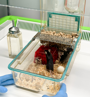
On account of their biological and genetic similarity to humans, mice are often deployed in research. With a knockout mouse, researchers literally knock out a certain gene or several genes. These genetically modified animals subsequently help them to examine a certain disease model. For the research on Fabry disease, these are knockout mice where the gene responsible for producing α-GAL A has been deactivated.
For the next step, Lorenz and Tolstik are planning to test the diagnostic method in human samples. If the method continues to prove its worth, it may in principle be used for other similar illnesses too, potentially in combination with further imaging procedures. The researchers at ISAS are thus building up further expertise in this vital new area where imaging procedures are used to find novel biomarkers for cardiovascular diseases. Collectively, these diseases kill around 18 million people annually around the world – more than every other cause of death. As non-destructive tools that provide an insight into the chemical composition of tissue and fluids, Raman and CARS spectroscopy may take on a key role in cardiovascular research.
(Ute Eberle)