Dortmund, 8th August 2024
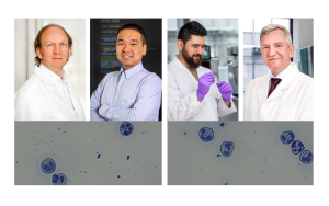
When pathogens attack our brain or spine, they're on the scene right away: microglial cells are a type of immune cell found in the central nervous system. They can play a key role in the development of neurodegenerative diseases such as Alzheimer's disease, Parkinson's disease or multiple sclerosis. Microglial cells are known for their ability to exhibit different phenotypes (observable characteristics) despite having an identical genetic blueprint. This heterogeneity at RNA and protein level has been known for a long time. But now researchers from the Justus Liebig University Giessen, Imperial College London and ISAS have been able to show for the first time that this heterogeneity also exists at lipid and, therefore, metabolic level for single cells. To do this, the scientists recorded the lipid signatures of single microglial cells: their unique, "fatty" signature. Until now it had only been possible to observe heterogeneity in cell cultures. The authors presented their research findings in the journal Analytical Chemistry.
Lipids are a molecular all-rounder: they form part of cell membranes, play an important role in a person's hormonal balance and provide cells with energy. Researchers describe the quantity, type and chemical structure of all the lipids in a particular area, such as a tissue or blood sample, using the lipid signature. They can use this unique signature to identify single cells and deduce how a particular cell metabolises the nutrients that are made available to it. So, the lipid signature helps to clarify which metabolic pathways may be affected by conditions such as Parkinson's disease, tumours or heart attacks. These diseases are highly heterogeneous, not all cells are affected equally and they have very different lipid signatures as a result.
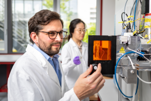
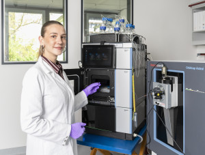
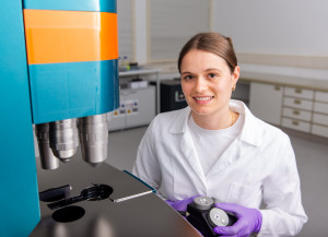
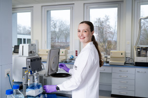
The team around Professor Dr-Ing. Sven Heiles, head of the Lipidomics junior research group, combines complementary techniques such as imaging mass spectrometry (MALDI: matrix-assisted laser desorption/ionisation) and light or fluorescence microscopy for their analyses. The photo shows Heiles (left) and doctoral student Chiahsin Chi at the MALDI mass spectrometer.
© ISAS / Hannes Woidich
The researchers used atmospheric pressure matrix-assisted laser desorption/ionization mass spectrometry (AP MALDI-MSI, see infobox) to decipher the signature of the microglial cells. Firstly, the scientists used laser beams to break the samples down into their molecular components so they could determine their type and frequency. Then they transformed this information into an image by means of software, which they could then use to determine the lipid signature and spatial distribution.
Better lasers allow for a higher resolution
To be able to detect the lipid signature of the minuscule cells involved, the authors first had to improve the local resolution of the chemical analysis. They used a specially made ionisation source , adjusted the laser energy and optimised the distance between the MS inlet capillary and the laser focal point. One single microglial cell has a diameter of 45 µm. Thanks to these optimisations, the researchers were ultimately able to maintain the signal intensity and record more than 100 different species of lipid per pixel.
AP MALDI-MSI
Atmospheric pressure matrix-assisted laser desorption/ionization mass spectrometry is a mass spectrometry imaging procedure. AP MALDI-MSI is especially suitable for analysing small biomolecules such as metabolites and lipids. A sample, such as a microglial cell in the case of the specified publication is exposed to a laser beam. This causes a fraction of the ablated sample material to desorb and ionise. A mass spectrometer can be used to detect the escaping molecular ions. The masses of the molecular ions are determined during this process, like on a scale. The measurements are usually accurate enough that conclusions can be drawn about the elemental composition and, thus, the type of biomolecule. The laser irradiation of the sample is repeated in the lab point by point. The researchers then obtain specialised software distribution images of the individual molecular ions..
Tell-tale fats point to inflammation
The researchers studied microglial cells in order to test their method on single cells. The authors simulated a bacterial inflammation and compared untreated cells against those stimulated with lipopolysaccharides (LPS). LPS is a molecule that is present in bacterial membranes. Treating cells with LPS therefore suggests an infection to them. The cells react to the supposed bacterial attack with an immune response designed to fight off the fictitious attackers. During this process, however, single cells "wear out" and can no longer handle the energy that is generated. These subgroups of cells store this excess energy in the form of triglycerides in lipid droplets. Triglycerides are lipids used by an organism to store energy. They can be found in lipid droplets or fat tissue, for example. The scientists were able to use the AP MALDI-MSI method that has been developed to distinguish the still active cells from the population of "worn out" cells. This allowed the researchers to show the heterogeneity of single microglial cells within a cell population at lipid level for the first time.
Article Recommendation
Müller, M.A., Zweig, N., Spengler, B., Weinert, M., Heiles, S.
(2023) Lipid Signatures and Inter-Cellular Heterogeneity of Naive and Lipopolysaccharide-Stimulated Human Microglia-like Cells. Analytical Chemistry, 95, 11672 – 11679. https://doi.org/10.1021/acs.analchem.3c01533.
Optimised treatment plans: The lipid signature could help
The Lipidomics junior research group at ISAS is collaborating with scientists from University Hospital Essen who are conducting research into skin cancer. Together, they want to use the lipid signature to determine whether heterogeneous behaviour – in this case, the aggressiveness of melanoma cells – depends on the lipid metabolism. In the long term, the ISAS researchers want to shed more light on how different cell types interact in living organisms. In future, the lipid signature could help to detect diseases such as skin cancer in diagnostic tissue – and, together with partners from clinical practice, to optimise treatments.
(Luisa Becher)