Dortmund, 28th March 2024
Large devices such as various mass spectrometers and microscopes, minor technical equipment, fume cupboards, refrigerators and freezers at minus 80 degrees Celsius – this is only a brief taste of all the technology for which ISAS needs electricity. In addition, there are other areas outside the laboratories which similarly require electrical power to operate. At the ISAS City location alone, electricity consumption amounted to 743,360 kilowatt hours in 2020. This is the same amount consumed on average by approximately 150 households of three or more persons in 2020.
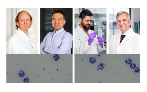
"The more highly developed the technology, the greater the information output. This results in increasing volumes of data but unfortunately also in more computing power being required to process it,” says Prof Dr Matthias Gunzer, head of the Biospectroscopy department at ISAS and Director of the Institute for Experimental Immunology and Imaging at University Hospital Essen. In order to drive ISAS forward in terms of sustainability, the institute had put a cogeneration unit into operation in 2019, set everything in motion for a photovoltaic system and decided to use liquefied gas. But do these measures alone suffice in the interests of performing climate-friendly research that is fit for the future? Gunzer’s answer is this: “It is also important to reduce the energy consumption of the technologies used in research. But at the same time, we would still like to increase their performance.” He went on to say that what initially sounds like a contradiction in terms can be implemented with clever planning and actually involves fascinating research and development work.

© Flaticon / ISAS
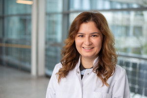
Prof Dr Matthias Gunzer heads the Biospectroscopy department and the Biofluorescence research group at ISAS. He is Director of the Institute for Experimental Immunology and Imaging at University Hospital Essen.
© ISAS / Hannes Woidich
Accurate image analysis despite low energy consumption
An example from the area of imaging makes one thing clear: technical progress goes hand in hand with ultra-high-resolution microscope images that have a high information content. These images generate large quantities of data. Storing and making this data available uses a lot of energy. In addition, analysis of the data using artificial intelligence (AI) requires substantial computing power, which in turn causes high power consumption. For this reason, AI specialists at ISAS are working towards reducing the energy consumed by data storage while still increasing the analysis quality of the images. To this end, they are first developing methods that make it possible to compress the data without losing key information. Less energy is consumed storing smaller files than larger ones. “We are also developing new software that extracts the maximum amount of image information from a kilowatt hour of electricity for the analysis calculations and, despite this low energy consumption, facilitates even more accurate image analyses than before,” adds Dr Jianxu Chen, head of the AMBIOM – Analysis of Microscopic BIOMedical Images – research group. But not only the processing and analysis of data play a role in reducing electricity consumption. For the ISAS researchers, what happens beforehand during microscopy work in the laboratory is crucial too.
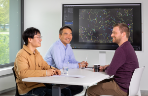
Together with their research group leader Dr Jianxu Chen (centre), doctoral candidates Yu Zhou (left) and Justin Sonneck are developing various AI-based tools for green microscopy.
© ISAS/Hannes Woidich
ComplexEye: Thirty times less energy than conventional microscopy
In order to track individual cell movements and cell shapes in real time, researchers at University Hospital Essen and ISAS have developed the ComplexEye. The prototype brings together in a single measuring device 16 microscopes (96 are planned for the future) that can take images simultaneously over a certain period of time of immune cells such as neutrophil granulocytes, for example. The researchers then combine these images into video sequences (so-called movies) of hundreds of individual migrating immune cells to create a time-lapse video.
“Although it generates more images in a shorter time than conventional microscopes, the ComplexEye currently consumes around 30 times less energy than a conventional system for the same amount of information,” Gunzer explains.
Immune cells are constantly searching the body for infectious intruders or incipient malignant diseases. However, migrating immune cells can themselves cause damage as well. For example, infiltration of growing tumours with neutrophils is associated with a poor prognosis for patients. The ComplexEye makes it possible to achieve a high throughput analysis of the migration of immune cells and provides important information that researchers were previously not able to gather. For example, the new microscope could help discover new kinds of active agent for cancer treatment, the effectiveness of which is based on stopping neutrophils migrating into tumours.
In order to find out how existing pharmaceutical active ingredients influence the migration of neutrophil granulocytes, the Essen-based researchers associated the samples with different substances via the Lead Discovery Center, Dortmund, in each case. For the subsequent analysis of the immune cells, the Dortmund-based AI specialists programmed a tailored application in 2022. “We developed a software based on various methods of artificial intelligence because common computer programmes for biomedical research reach their limits with this large number of movies,” says Chen. This information gathered using ComplexEye and evaluated with the help of AI also makes new means of diagnostics possible – for example, to detect sepsis (blood poisoning) earlier and thus be better able to treat it.
Article Recommendation
Cibir, Z., Hassel, J., Sonneck, J., Kowitz, L., Beer, A., Kraus, A., Hallekamp, G., Rosenkranz, M., Raffelberg, P., Olfen, S., Smilowski, K., Burkard, R., Helfrich, I., Tuz, A. A., Singh, V., Ghosh, S., Sickmann, A., Klebl, A-K., Eickhoff, J. E., Klebl, B., Seidl, K., Chen, J., Grabmaier, A., Viga, R., Gunzer, M. (2023). ComplexEye: a multi-lens array microscope for high-throughput embedded immune cell migration analysis. Nat Commun 14, 8103. https://doi.org/10.1038/s41467-023-43765-3.
The MSCoreSys associated junior research group AMBIOM – Analysis of Microscopic BiOMedical Images is funded by the Federal Ministry of Education and Research (Bundesministerium für Bildung und Forschung, BMBF) under the funding reference 161L0272.