Skip to content

Red Alert: Students Research the Immune System at Girls' Day

Dortmund, 8th May 2024

For half a day at Girls' Day 2024, twelve schoolgirls were able to find out in their own experiments how bacteria can be hunted down and kept in check, why organs become transparent with the help of cinnamic acid ethyl ester, and what can be seen under the microscope during a heart attack.

Together with PhD students Antonia Fecke and Luisa Speicher, who accompanied the event as scientific guides, the seventh to ninth graders gained practical insights into the immune system – and learned a lot about bacterial and sterile inflammation. Under the guidance of Dr Christina Sengstock and Dr Christiane Stiller, the pupils learned how to multiply bacterial cultures in the laboratory. They also investigated the effect of silver acetate on Escherichia coli. With Prof. Dr Anika Grüneboom and Luisa Röbisch, everything revolved around the light sheet fluorescence microscope. Before they treated organs with cinnamic acid ethyl ester – a component of cinnamon flavour – and analysed pre-treated samples, the participants had conducted an experiment with glass beads to gain a better understanding of the procedure for optical clearing.

The Communications team coordinated the Girls' Day project at ISAS in 2024, and this time received active support with the organisation and implementation from intern Clara Manthey (student at TU Dortmund University). In the run-up to the event, Luisa Becher, also a student at TU Dortmund University, had already played a key role in the conception and preparation.

Im Bild pipettiert eine der Schülerinnen Nährmedium auf eine Agarplatte. Die Platte ist rechts im Bild nochmal detailliert zu sehen. In the picture, one of the students is pipetting culture medium onto an agar plate. A plate can be seen again in detail on the right of the picture.

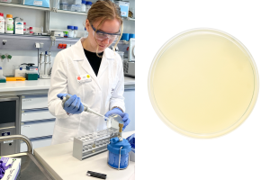
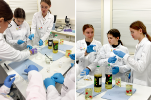
In the bacteria lab, Jule (13) is practising how to use a pipette. The work is taking place next to a Bunsen burner the whole time. It forms a sterile circle around its flame, in which it is possible to work in a sterile environment. Within this sterile zone, Jule is training how to pipette, first with water and then with a prepared Luria-Bertani medium. The latter is a nutrient medium for cultivating bacteria. For this, the 8th grader has already placed a new sterile plastic tip onto the pipette before she applies the medium to the prepared agar plate. The aim of this experiment is for Jule and the other pupils to learn how to handle bacterial cultures (E. coli) and how to multiply them to continue working with them afterwards.

© ISAS (links) istockphoto/grebcha (rechts)

Das BIld zeigt zwei Schülerinnen, wie sie unter Anleitung von Dr. Christina Sengstock Nährmedium auf eine Agarplatte pipettieren. Rechts daneben ist eine Agarplatte mit Hemmhöfen um zwei mit Silberacetat behandelte Areale zu erkennen. The picture shows two students pipetting culture medium onto an agar plate under the guidance of Dr. Christina Sengstock. To the right is an agar plate with inhibition zones around two areas treated with silver acetate.

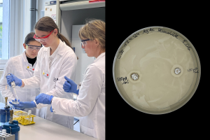
Once the students have familiarised themselves with pipetting, they move on to the “inhibition zone experiment”. Dr Christina Sengstock (on the right in the picture) is standing by to help them. Sara (14, photo: centre) has already labelled the Petri dish with the culture medium. She is now holding an agar plate and a pipette in her hands. She uses the pipette to drip Luria-Bertani medium onto the plate. The student makes sure that she uses the exact amount of liquid required. The girls have previously taken the liquid from a test tube using the pipette. They will then spread the medium on the plate by spreading it out with a glass spatula they had bent themselves. Sophie (13) is watching attentively before pipetting herself next. The whole exercise serves as preparation for the following experiment. For this, the pupils will make holes into the agar on the plate. They will then pipette silver acetate into the punched-out area. Agar plates with E. coli-cultures are used in real daily life in the laboratories. The silver ions have an antimicrobial effect – they inhibit the growth of bacteria. That is why inhibition zones around the area treated with silver acetate can later be seen with the bare eye.

© ISAS

Links im Bild sind zwei Teilnehmerinnen zu sehen, wie sie ein Becherglas mit Speiseöl füllen. Rechts im Bild ist das gefüllte Glas und das darin enthaltene Acryl-Herz zu erkennen. The picture on the left shows two participants filling a beaker with cooking oil. The filled glass and the acrylic heart it contains can be seen on the right.

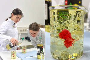
The young researchers have moved from the bacteria lab to one of the microscopy labs. To understand how organs become transparent for the analysis under the light sheet fluorescence microscope, the students have been given cups with glass pearls. They are part of an experiment conducted by Prof. Dr Anika Grüneboom and Luisa Röbisch. The two have chosen this experiment to illustrate the principle of optical clearing. What Sophie (left) and Sara cannot see at first because the glass beads do not let any light through, and the beaker appears opaque: There are small red plastic hearts between the glass beads. The pupils are slowly pouring cooking oil into the beaker. The oil displaces the air between the glass beads. The similar refractive index of glass and oil ensures that the hearts in the beakers become visible. 

© ISAS

Das Bild zeigt die Teilnehmerinnen bei der Arbeit mit den Organproben. In den Händen halten sie kleine Glasgefäße mit Ethanollösung und den Organen. The picture shows the participants working with the organ samples. They are holding small glass containers with ethanol solution and the organs.

Sara (right) and Sophie are working with organ samples. They are carefully preparing the delicate samples for later use under the light sheet fluorescence microscope. They are using tweezers to pick up the hearts and thymus glands of mice, and take them out of an ethanol solution. Prof Dr Anika Grüneboom and Luisa Röbisch have prepared samples of the heart, bowel, and thymus for the experiment in advance and placed them in an ethanol solution. This alcohol solution is used to dehydrate the samples. The students are carefully placing the samples in small containers filled with ethyl cinnamate before they will seal the jar. The cinnamic acid ethyl ester is part of Prof. Dr Anika Grüneboom patented clearing process. (Editor's note: The samples were already available; no organ removals were carried out for the event).

© ISAS

Links im Bild schaut sich eine Schülerin eine Organprobe an und hält sie gegen das Licht. Rechts ist eine Lightsheetaufnahme des Herzenz zu sehen, nachdem sie am Computer zusammengesetzt wurde. n the left, a student looks at an organ sample and holds it up to the light. On the right is a lightsheet image of the heart after it has been assembled on the computer.

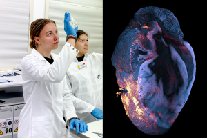
Jule (left) is taking a close look at the bowel. She can already see that it is transparent. The lab is darkened with roller blinds because the students will next go to the lightsheet fluorescence microscope. Because clearing some of the organs takes several days, biotechnologist Luisa Röbisch had prepared some samples for Girls' Day a few days before the event. Immunologist Prof. Dr Anika Grüneboom will later place a transparent heart under the light sheet microscope. The device fans out the dot-shaped laser beam like a sheet of paper. The thin sheet of light created in this way illuminates each individual layer of the sample. Because the samples are transparent, the laser can penetrate them almost unhindered – and take an image of each layer. At Girls' Day, Grüneboom will later discuss the individual images of a heart after a heart attack with the students. And she will then assemble these images into a 3D model on the computer. (Editor's note: The samples were already available; no organ removals were carried out for the event).

© ISAS

Das Bild zeigt einen Teil der Schülerinnen zusammen mit Luisa Röbisch, Dr. Anika Grüneboom und Dr. Christiane Stiller, während sie im Labor stehen und in die Kamera schauen. Auf der rechten Seite ist eine Aufnahme des Herzgewebes mit dem Konfokal Mikroskop zu sehen. The picture shows some of the students together with Luisa Röbisch, Dr Anika Grüneboom and Dr Christiane Stiller as they stand in the lab and look into the camera. On the right is an image of the heart tissue taken with the confocal microscope.

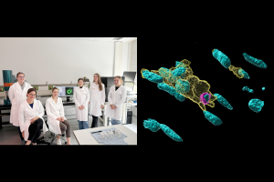
The last part of the programme is about how pathogens – in this case E. coli bacteria – multiply and look on the agar plates after a few days. Another topic: immune cells such as macrophages which exaggerate with “cleaning up” after a heart attack and therefore damage the body (sterile inflammation). At this part of the Girls’ Day, the researchers have also shown images that they had taken under a confocal microscope. They revealed a section of the heart tissue with an accumulation of macrophages. Afterwards, Dr Christiane Stiller (from left to right), Luisa Röbisch and Prof. Dr Anika Grüneboom are answering the students’ questions about their day-to-day work at ISAS. 

© ISAS

Gruppenfoto aller Teilnehmerinnen und Mitwirkenden im ISAS Foyer. Im Hintergrund buchstabieren große Ballons “I LOVE SCIENCE“. Group photo of all participants and contributors in the ISAS foyer. In the background, large balloons spell out “I LOVE SCIENCE“.

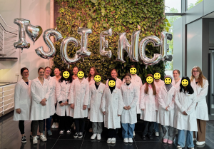
So many bright and curious minds took part in the Girls' Day event at ISAS. At the end of the day, the twelve students gave positive feedback on the day at the institute. And who knows, maybe the photo also shows the next generation of women in research? In any case, the motto “I love science” applies to all those present. Luisa Röbisch, Prof. Dr Anika Grüneboom, Dr Christiane Stiller, Cheyenne Peters (Communications team), Clara Manthey (intern in the Communications team), Luisa Speicher and Antonia Fecke (from left to right) also had a lot of fun carrying out the experiments and presenting their work. (Editor's note: Dr Christina Sengstock and Sara Rebein are missing from the photo).

© ISAS

Share

Further articles

2nd May 2024

Hydrogen Sulfide: The Surprising Molecule That Regulates Life's Vital Functions & Fights Aging

Hydrogen sulfide is considered highly toxic. Nevertheless, the foul-smelling gas fulfills many vital functions in our cells. As a gasotransmitter, for example, it can transmit signals within and between cells. But hydrogen sulfide also plays an important role in the oxygen supply in the blood – as researchers led by Dr habil Miloš Filipović at ISAS recently discovered.

Porträt Dr. habil. Miloš Filipović.
28th March 2024

New “Green” Microscopy: Less Electricity, but More Information on Immune Cells in Return

Advanced technologies, such as high-resolution microscopes, produce large amounts of data. And these again consume large amounts of electricity. In addition, there are refrigerators for samples, fume cupboards and small technical devices. While ISAS is upgrading to become greener, researchers at the institute are already working on methods to make microscopy more energy-efficient in general.

Das Bild zeigt eine schematische Darstellung der Datenverarbeitung in der Mikroskopie.
13th March 2024

Cirrhosis of the Liver: Migrating Immune Cells Act as an Early Warning System

The life expectancy of patients with liver cirrhosis depends crucially on the occurrence of disease-associated complications, such as infections. Until now, however, an ability to predict these at an early stage has been lacking. A problem, often hindering doctors from administering antibiotics or even performing a liver transplant in time. Researchers at ISAS led by Prof Dr Matthias Gunzer therefore investigated the question: Could the mobility of certain immune cells be the decisive indicator of an impending deterioration in health?

27th February 2024

3 Questions for… Dr Christopher Nelke

As a participant in the Clinician Scientist programme and physician at the Clinic for Neurology at Düsseldorf University Hospital (Universitätsklinikum Düsseldorf, UKD), Dr Christopher Nelke researches neuromuscular diseases. In this interview, he talks about his two-week guest stay at ISAS and the challenges that arise between the hospital bed and research.

Das Bild zeigt Dr. Christopher Nelke im Labor. In den Händen hält er eine Probe. The picture shows Dr Christopher Nelke in the laboratory. He is holding a sample in his hands.
20th February 2024

SARS-CoV-2: The Very Latest Methods Clarify the Active Agents and the Mechanism of Action of Ancient Self-Medications

Prophylactic, soothing or even healing agents, mostly natural substances, have been known to natural medicine since ancient times. But what about viral infections? Could infusions made from sage or perilla also be used against SARS-CoV-2 infections -as a prevention or an aid to healing? An interdisciplinary team of researchers led by Prof Dr Mirko Trilling from the Faculty of Medicine at the University of Duisburg-Essen (UDE) and scientists at ISAS investigated these questions during the coronavirus pandemic.

Das Bild zeigt Prof. Dr. Mirko Trilling mit verschränkten Armen, an einer Wand lehnend. The picture shows Prof Dr Mirko Trilling with his arms folded, leaning against a wall.
7th February 2024

A Long Sought-After Combination Method in Mass Spectrometry

Researchers who analyse complex samples using mass spectrometers are often faced with the challenge that the substances they contain are fundamentally different. Some are chemically polar, others are non-polar. Until now, this has required two complex separate analyses. But a researcher at ISAS has developed a method with which even less polar substances can be included in a standard mass spectrometric analysis for polar biological substances.

Daniel Foest steht im Labor und hält ein Papier mit einer Leberprobe, die er am Massenspektrometer untersucht.
12th January 2024

“My research is literally hard work"

Darleen Hüser is looking for the immuno-cellular fingerprint of rheumatoid arthritis. In this interview, the doctoral student reveals her razor-sharp research and why she needs different microscopes.

Das Porträt zeigt ISAS-Doktorandin Darleen Hüser aus der Arbeitsgruppe Bioimaging.
21st December 2023

Science Slam: Humorous Science Communication Is Fun for Everyone

Talking lab equipment, artificial intelligence and expertise from the North Pole - this colourful mix of topics characterised the latest Science Slam at the institute. Four ISAS employees demonstrated with their specialist knowledge and plenty of humour how science communication can be fun for everyone involved.

Luisa Becher fotografiert die vier Teilnehmenden des ISAS Science Slam.
20th December 2023

The Art of Balancing: Accuracy in Image Analysis

What challenges in analysing microscopic images can be mastered with artificial intelligence if the latter is incorporated at an early stage? Why should researchers think about the target metrics of image analysis when planning their experiment? Dr Jianxu Chen has now put together his tips, recently published in the journal Nature Methods, as a kind of checklist for researchers.

Die Abbildung zeigt eine Wage und symbolisiert das Gleichgewicht zwischen Analyse und Genauigkeit bei der Validierung von biomedizinischen Aufnahmen.