Photos
-
-

-
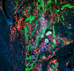
Rheumatoid Arthritis: Murine Knee Joint (II)
Download fileInfiltration of immune cells (red) and inflammation-driven angiogenesis of blood vessels (green and white) in a murine knee joint during rheumatoid arthritis. The image was taken using confocal laser scanning microscopy.
-

-
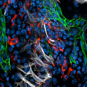
Rheumatoid Arthritis: Murine Knee Joint (I)
Download fileInfiltration of immune cells (red) and inflammation-driven angiogenesis of blood vessels (green and white) in a murine knee joint during rheumatoid arthritis. The image was taken using confocal laser scanning microscopy.
-
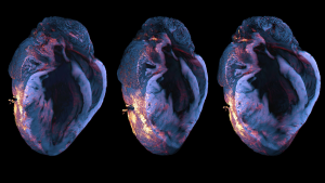
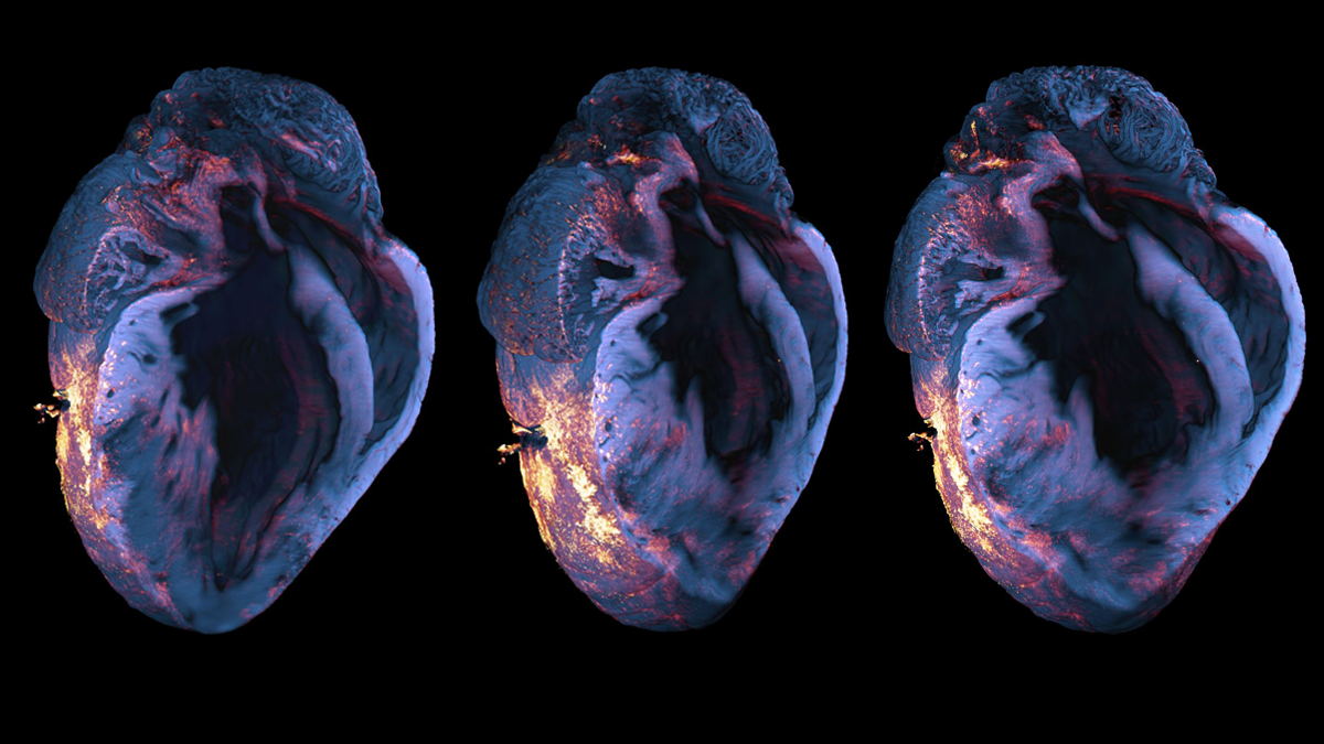
Myocardial Infarction: Immune Cell Infiltration
Download fileLight sheet fluorescence microscopy of immune cell infiltration (red-orange) in a murine heart (blue) after myocardial infarction. The image was taken using confocal laser scanning microscopy.
-

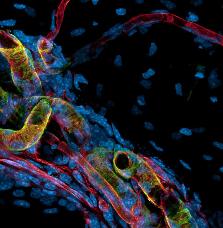
Periosteum: Blood Vessels
Download fileTo ensure bone homeostasis, the bone surface – the periosteum – and the cortical bone are supplied by different types of blood vessels, such as fine capillaries (red) and large arteries (green and red). The image was taken using confocal laser scanning microscopy.
-
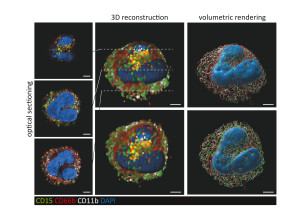
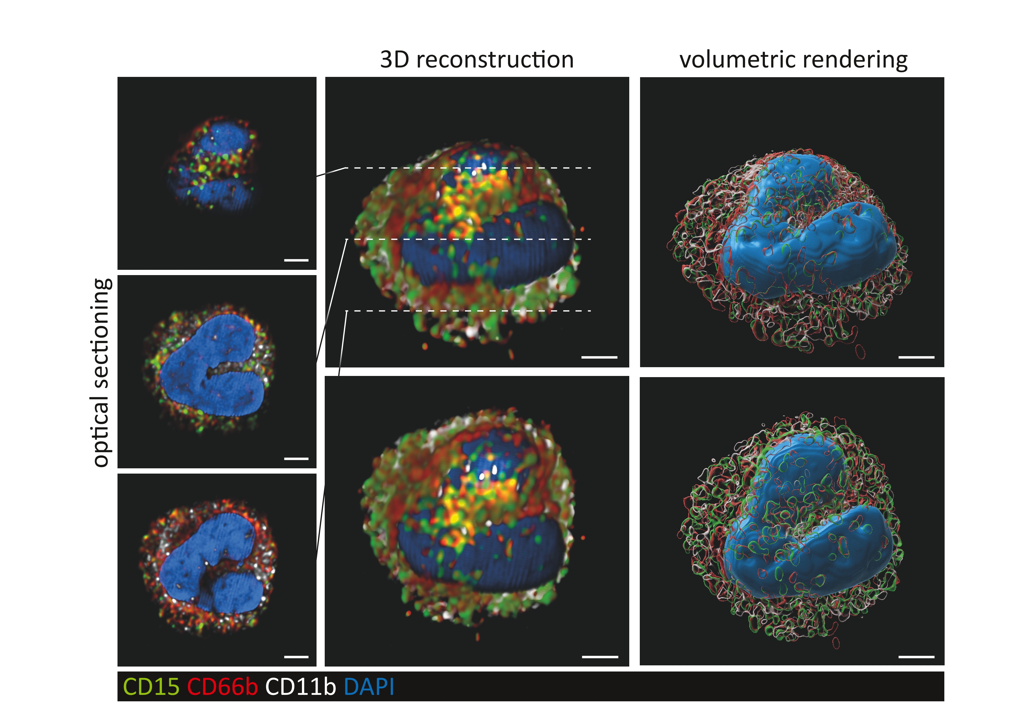
Human Neutrophil Granulocyte
Download file3D Reconstruction of a Human Neutrophil Granulocyte. The image was taken using confocal laser scanning microscopy.
-

